Download

1 / 50
500 likes | 623 Vues
The muscular system is essential for body movement and involves three muscle types: skeletal, cardiac, and smooth. Skeletal muscles are striated, voluntary, and attached by tendons to bones. Cardiac muscles are striated and involuntary, found only in the heart. Smooth muscles are non-striated and also involuntary, located in hollow organs. Muscles contract through the sliding filament theory, using ATP for energy. Understanding muscle anatomy, including connective tissues and nerve stimulation, is crucial for grasping how movements are generated and regulated in the body.

E N D
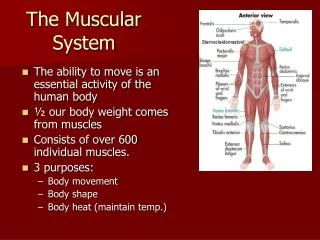
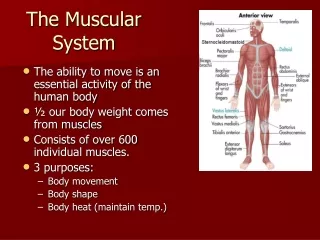
The Muscular System • Muscles are responsible for all types of body movement • Three basic muscle types are found in the body • Skeletal muscle • Cardiac muscle • Smooth muscle
Characteristics of Muscles • Muscle cells are elongated (muscle cell = muscle fiber) • Contraction of muscles is due to the movement of microfilaments • All muscles share some terminology • Prefix myo refers to muscle • Prefix mys refers to muscle • Prefix sarco refers to flesh
Skeletal Muscle Characteristics • Most are attached by tendons to bones • Cells are multinucleate • Striated – have visible banding • Voluntary – subject to conscious control • Cells are surrounded and bundled by connective tissue
Connective Tissue Wrappings of Skeletal Muscle • Endomysium – around single muscle fiber • Perimysium – around a fascicle (bundle) of fibers Figure 6.1
Connective Tissue Wrappings of Skeletal Muscle • Epimysium – covers the entire skeletal muscle • Fascia – on the outside of the epimysium Figure 6.1
Skeletal Muscle Attachments • Epimysium blends into a connective tissue attachment • Tendon – cord-like structure • Aponeuroses – sheet-like structure • Sites of muscle attachment • Bones • Cartilages • Connective tissue coverings
Smooth Muscle Characteristics • Has no striations • Spindle-shaped cells • Single nucleus • Involuntary – no conscious control • Found mainly in the walls of hollow organs Figure 6.2a
Cardiac Muscle Characteristics • Has striations • Usually has a single nucleus • Joined to another muscle cell at an intercalated disc • Involuntary • Found only in the heart Figure 6.2b
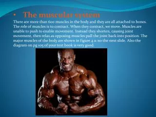
Function of Muscles • Produce movement • Maintain posture • Stabilize joints • Produce heat
Microscopic Anatomy of Skeletal Muscle • Cells are multinucleate • Nuclei are just beneath the sarcolemma (specialized plasma/cell membrane) Figure 6.3a
Microscopic Anatomy of Skeletal Muscle • Sarcoplasmic reticulum – specialized smooth endoplasmic reticulum that stores calcium Figure 6.3a
Microscopic Anatomy of Skeletal Muscle • Myofibril • Organelle made of bundles of myofilaments • Myofibrils are aligned to show distinct bands • I band = light band • A band = dark band Figure 6.3b
Microscopic Anatomy of Skeletal Muscle • Sarcomere • Contractile unit of a muscle fiber Figure 6.3b
Microscopic Anatomy of Skeletal Muscle • Organization of the sarcomere • Thick filaments = myosin filaments • Composed of the protein myosin which has heads (extensions, or cross bridges) • Has ATPase enzymes Figure 6.3c
Microscopic Anatomy of Skeletal Muscle • Organization of the sarcomere • Thin filaments = actin filaments • Composed of the protein actin Figure 6.3c
Microscopic Anatomy of Skeletal Muscle • Myosin and actin overlap somewhat Figure 6.3d
Nerve Stimulus to Muscles • Skeletal muscles must be stimulated by a nerve to contract • Motor unit • One neuron • Muscle cells stimulated by that neuron Figure 6.4a
Nerve Stimulus to Muscles • Neuromuscular junctions – association site of nerve and muscle Figure 6.5b
Nerve Stimulus to Muscles • Synaptic cleft – gap between nerve and muscle • Nerve and muscle do not make contact • Area between nerve and muscle is filled with interstitial fluid Figure 6.5b
Transmission of Nerve Impulse to Muscle • Neurotransmitter – chemical released by nerve upon arrival of nerve impulse • The neurotransmitter for skeletal muscle is acetylcholine • Neurotransmitter attaches to receptors on the sarcolemma • Sarcolemma becomes permeable to sodium (Na+)
Transmission of Nerve Impulse to Muscle • Sodium rushing into the cell generates an action potential • Once started, muscle contraction cannot be stopped muscle contraction animation
The Sliding Filament Theory of Muscle Contraction • Activation by nerve causes myosin heads (crossbridges) to attach to binding sites on the thin filament • Myosin heads then bind to the next site of the thin filament Figure 6.7
The Sliding Filament Theory of Muscle Contraction • This continued action causes a sliding of the myosin along the actin • The result is that the muscle is shortened (contracted) Video about cross bridge cycle (sliding filament process) by Pearson Figure 6.7
The Sliding Filament Theory Figure 6.8
Muscle Response to Strong Stimuli • Muscle force depends upon the number of fibers stimulated • More fibers contracting results in greater muscle tension • Muscles can continue to contract unless they run out of energy
Energy for Muscle Contraction • Initially, muscles use stored ATP for energy • Bonds of ATP are broken to release energy • Only 4-6 seconds worth of ATP is stored by muscles • After this initial time, other pathways must be utilized to produce ATP
Energy for Muscle Contraction • Direct phosphorylation • Muscle cells contain creatine phosphate (CP) • CP is a high-energy molecule • After ATP is depleted, ADP is left • CP transfers energy to ADP, to regenerate ATP • CP supplies are exhausted in about 20 seconds Figure 6.10a
Energy for Muscle Contraction • Aerobic Respiration • Series of metabolic pathways that occur in the mitochondria • Glucose is broken down to carbon dioxide and water, releasing energy • This is a slower reaction that requires continuous oxygen Figure 6.10b
Energy for Muscle Contraction • Anaerobic glycolysis • Reaction that breaks down glucose without oxygen • Glucose is broken down to pyruvic acid to produce some ATP • Pyruvic acid is converted to lactic acid Figure 6.10c
Energy for Muscle Contraction • Anaerobic glycolysis (continued) • This reaction is not as efficient, but is fast • Huge amounts of glucose are needed • Lactic acid produces muscle fatigue Figure 6.10c
Muscle Fatigue and Oxygen Debt • When a muscle is fatigued, it is unable to contract • The common reason for muscle fatigue is oxygen debt • Oxygen must be “repaid” to tissue to remove oxygen debt • Oxygen is required to get rid of accumulated lactic acid • Increasing acidity (from lactic acid) and lack of ATP causes the muscle to contract less
Muscles and Body Movements • Movement is attained due to a muscle moving an attached bone Figure 6.12
Muscles and Body Movements • Muscles are attached to at least two points • Origin – attachment to a moveable bone • Insertion – attachment to an immovable bone Figure 6.12
Effects of Exercise on Muscle • Results of increased muscle use • Increase in muscle size • Increase in muscle strength • Increase in muscle efficiency • Muscle becomes more fatigue resistant
Types of Ordinary Body Movements • Flexion • Extension • Rotation • Abduction • Circumduction
Body Movements Figure 6.13a–c
Body Movements Figure 6.13d
Special Movements • Dorsiflexion • Plantar flexion • Inversion • Eversion • Supination • Pronation • Opposition
Types of Muscles • Prime mover – muscle with the major responsibility for a certain movement • Antagonist – muscle that opposes or reverses a prime mover • Synergist – muscle that aids a prime mover in a movement and helps prevent rotation • Fixator – stabilizes the origin of a prime mover
Naming of Skeletal Muscles • Direction of muscle fibers • Example: rectus (straight) • Relative size of the muscle • Example: maximus (largest)
Naming of Skeletal Muscles • Location of the muscle • Example: many muscles are named for bones (e.g., temporalis) • Number of origins • Example: triceps (three heads)
Naming of Skeletal Muscles • Location of the muscle’s origin and insertion • Example: sterno (on the sternum) • Shape of the muscle • Example: deltoid (triangular) • Action of the muscle • Example: flexor and extensor (flexes or extends a bone)
Head and Neck Muscles Figure 6.15
Trunk Muscles Figure 6.16
Deep Trunk and Arm Muscles Figure 6.17
Muscles of the Pelvis, Hip, and Thigh Figure 6.19c
Muscles of the Lower Leg Figure 6.20
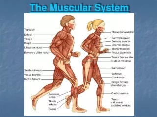
Superficial Muscles: Anterior Figure 6.21
Superficial Muscles: Posterior Figure 6.22