Download
1 / 15
150 likes | 297 Vues
Ion Beam Analysis Dolly Langa Physics Department, University of Pretoria, South Africa Blane Lomberg Physics Department, University of the Western Cape, South Africa Project Supervisor: Prof A.P. Kobzev Frank Laboratory of Neutron Physics, Joint Institute for Nuclear Research,

E N D
Ion Beam Analysis Dolly Langa Physics Department, University of Pretoria, South Africa Blane Lomberg Physics Department, University of the Western Cape, South Africa Project Supervisor: Prof A.P. Kobzev Frank Laboratory of Neutron Physics, Joint Institute for Nuclear Research, Dubna, Russia
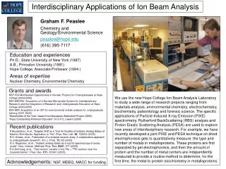
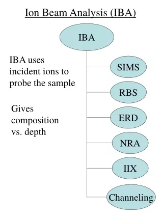
AIM OF PROJECT • Analysis of contents and depth distribution of different elements in the near surface layers of solids using • Rutherford Backscattering Spectrometry (RBS) • Elastic Recoil Detection (ERD) • Particle Induced X-ray Emission (PIXE)
OUTLINE • 1. AIM OF PROJECT • 2. VAN DER GRAAFF ACCELERATOR • 3. PRINCIPLE OF ION BEAM ANALYSIS USED • 4. RESULTS AND DISCUSSION • 5. CONCLUSION
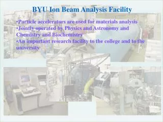
Van de Graaff Accelerator Parameters at JINR • Produces the beams of helium ions and protons with energy in regions 0.9-3.5 MeV • Helium intensity less than 10 A and proton intensity up to 30 A. • Energy spread less than 500 eV • The accelerator belt moves at 20 m/s • The accelerator is placed in a tank under pressure of 10 atmospheres of dry nitrogen. • The accelerator EG-5 has six beam lines.
PRINCIPLE OF ION BEAM ANALYSIS USED Conti.. Rutherford Backscattering Spectrometry (RBS)
RESULTS AND DISCUSSIONRBS spectrum for the sample with the Fe and Ti layers on Si substrate, with Ti layer containing Oxygen. Calibration: Calibration offset = 35.72 keV Energy per channel = 1.8782 keV/ch Thickness: Fe = 76 nm Ti = 62 nm Concentrations in Ti layer: Ti = 30 at % O = 70 at % EHe = 2.035 MeV = 100 = 1700 Fe Oxygen Ti Si Substrate
RBS spectrum for the sample with the Ge and Si multi-layers on Si substrate EHe = 1 MeV = 300 = 200 = 1700 Ge Si substrate Si
PRINCIPLE OF ION BEAM ANALYSIS USEDRutherford Backscattering Spectrometry (RBS) and Elastic Recoil Detection (ERD) setup
RBS and ERD spectra Thickness: (C) = 170 nm Thickness (O) = 20 nm Si = 26 at % Si = 70 at % H = 40 at % H = 20 at % C = 34 at % O = 10 at % EHe = 2.297 MeV = 750 = 750 = 300 Thickness (H) = 190 nm C O • EHe = 2.297 MeV • = 750 = 300 = 1350 Si
PRINCIPLE OF ION BEAM ANALYSIS USED Conti..Particle Induce X-ray Emission (PIXE)
CONCLUSION • These methods are non-destructive techniques to study materials • The used methods allow the determination of depth distribution and concentration from hydrogen to heavy elements. • The spectra calculations and model comparisons was executed in SIMNRA software tool, in which good agreement was achieved for RBS and ERD experiments. • Furthermore, the depth resolution is done near to few nm range for these methods. • The sensitivity for heavy elements is of the order 1014 atoms/cm2