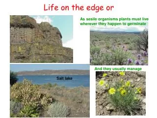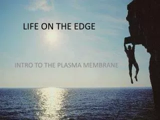
LIFE ON THE EDGE
620 likes | 822 Vues
LIFE ON THE EDGE. INTRO TO THE PLASMA MEMBRANE. MEMBRANE MODELS (from past to present). 1915: membranes isolated from RBC’s were analyzed & found to be made of lipids & proteins

LIFE ON THE EDGE
E N D
Presentation Transcript
LIFE ON THE EDGE INTRO TO THE PLASMA MEMBRANE
MEMBRANE MODELS (from past to present) • 1915: membranes isolated from RBC’s were analyzed & found to be made of lipids & proteins • 1925: scientists (Gorter & Grendel) suggested membrane was made of phospholipid bilayer. (hydrophobic parts are sheltered by the hydrophillic parts). Where are the proteins?
1935: scientists Davidson & Danielli suggested the membrane was like a sandwich. • Phospholipid bilayer b/w layers of proteins.
1950’s: came the invention of the electron microscope. • Pictures seemed to support the sandwich model • 1960’s: scientists had problems with model • Cells with different functions differed in structure and chemical make-up. • Proteins in membranes were not very soluble (they were amphipathic). If these proteins were the “bread” part of the sandwich the hydrophobic parts would be in an aqueous environment.
1972: Singer and Nicolson proposed the membrane proteins were dispersed & individually inserted into the phospholipid bilayer. • Only the hydrophillic portions are protruding Fluid Mosaic Model
Freeze fracture (method of preparing cells for the electron microscope)- confirmed Singer and Nicolson’s model.
Fluid mosaic model • Not static or rigid • Proteins & lipids drift back & forth (laterally) • Movement is rapid • Proteins move more slowly than lipids • Some move as directed (motor proteins attached) • Some don’t move (anchored)
The fluidity of a lipid bilayer depends on both its composition and its temperature • The temperature at which the membrane solidifies is dependent on the types of lipids in the membrane • Unsaturated lipids create a kink, preventing the fatty acids from packing together as tightly, thus decreasing the melting temperature (increasing the fluidity) of the membrane.
Unsaturated vs Saturated Fatty Acids in the Membrane • Plants, animals, & bacteria adapt to decreasing temperatures by increasing the proportion of unsaturated fatty acids in the membrane. • They decrease the proportion of unsaturated fatty acids in the membrane & increase the saturated fatty acid content in the membrane when temps are high.
The ability of some organisms to regulate the fluidity of their cell membranes by altering lipid composition is called homeoviscous adaptation.
NOW LET’S TALK CHOLESTEROL • Is amphipathic. • The hydrophobic steroid ring which is near the hydrocarbon tails of the phospholipids tend to immobilize the tails. • Restricts random movement so membrane doesn’t turn to mush. • Keeps lipids more separated so they don’t crystalize. • (temperature buffer)
How would you expect the saturation levels of membrane fatty acids to differ in plants adapted to cold environments & plants adapted to hot environments? • Plants in cold environments would probably have more unsaturated fatty acids in their membrane, since those remain fluid at lower temps. • Plants in hot environments would probably have more saturated fatty acids to so they would be closer to each other, causing the membrane to be less fluid. Being less fluid would help them stay intact at higher temperatures.
It’s the “mosiac” turn • Proteins - Over 50 different types • Different cells = different proteins • Proteins determine the function of the membrane therefore the function of the cell.
2 Major Types of Proteins • Integral: • Peripheral -many are transmembranes – amphimathic -exposed to both aqueous solutions (inside & outside of the cell) -not embedded in the lipid bilayer -they are appendages to integral proteins & are attached to the cytoskeleton
Integral Peripheral
Catalyze reactions inside the cell ENZYAMTATIC Bind cells together to make tissues INTRACELLULAR JOINING Allows cells to bind to each other or a substrate ATTACHMENT TO CYTOSKELETON & THE ECM Moves molecules in & out of the cell using passive or active passageways. TRANSPORT Convey signals from the outside of the cell to the inside; functions like a light switch. SIGNAL TRANSDUCTION Allow immune system to recognize what is self & not self RECOGNITION
CARBOHYDRATES OF THE MEMBRANE • Cell to Cell Recognition: • This comes in handy when in the embryonic stage • Basis for rejection of organ transplants by immune system. • 2 types • Glycolipids- carbohydrate bonded to a lipid • Glycoprotein- carbohydrate bonded to a protein • Function as markers- distinguish one cell from the other. • Ex: human blood types: each have variation in their carbohydrate markers.
How does the endoplasmic reticulum make the plasma membrane?
WHAT IS THE PLAMA MEMBRANE MADE OF? • LIPIDS • PHOSPHOLIPIDS (amphipathic) • CHOLESTEROL • PROTEINS • INTEGRAL PROTEINS (amphipathic) • PERIPHERAL PROTEINS • CARBOHYDRATES • GLYCOLIPIDS (carbohydrate and lipid) • GLYCOPROTEINS (carbohydrate and protein)
CONSTRUCTING A MODEL OF THE CELL MEMBRANE • Construct a model of the cell membrane using diagrams you have been provided in your text, during lecture, and in this lab. • You may use construction material provided (assorted food & non-food materials: assorted pasta, assorted cereals, glue, pipe cleaners, yarn, and more) or supply additional material on your own. • Make sure your model includes the following components: a. Phospholipid bilayer (heads & tails!) b. Integral proteins c. Peripheral proteins d. Glycolipids e. Carbohydrates f. Glycoproteins g. Cholesterol • Make a key to show what each item represents • Glue your cell membrane to cardboard • Answer the Summary Questions on the next slide.
How do molecules pass in and out of the plasma membrane? • If you (the molecule) are small and non-polar OR small & uncharged you can pass through the lipid bilayer. • The hydrophobic tails of the lipid bilayer impedes ions and polar molecules. • This includes glucose and water!!
Transport Proteins • Hydrophylic substances (some ions and polar molecules) pass through the membrane by way of the channel proteins • aquaporins facilitate the passage of water
Transport Proteins • Carrier proteins: these proteins change shape as they pass substances through. They are highly selective.
What mechanisms drive molecules across the membrane? • Passive Transport • Diffusion • Osmosis • Facilitated diffusion • Active Transport • Sodium Potassium Pump/Electrogenic pump • Cotransport • Exocytosis • Endocytosis
Diffusion • A substance will travel from where it is more concentrated to where it is less concentrated. • Substances travel down its concentration gradient. • No work needs to be done • No need for energy • It is a spontaneous process • Most travel of substances across is by diffusion • EX: oxygen transfer; CO2transfer
Osmosis • diffusion of water molecules • across a selectively permeable membrane • Movement is from areas of high potential (high free water concentration) and low solute concentration to areas of low potential (low free water concentration) & high solute concentration.
WATER MOLECULES CLUSTERED AROUND HYDROPHILIC MOLECULES ARE UNAVAILABLE TO TRAVEL THROUGH THE MEMBRANE.
Solutions of Osmosis • HYPERTONIC: • Has a higher solute concentration and a lower water potential compared to the solution on the other side of the membrane. • HYPOTONIC: • Has a lower solute concentration and a higher water potential than the solution on the other side of the membrane • ISOTONIC: • Have equal water potentials
Cells without cell walls • In a hypotonic environment, water enters cell and it swells and may burst • In a hypertonic environment, water leaves the cell and it shrinks
Turgor Pressure • most plant cells live in hypotonic environment • water moves into cells, pushing cell membrane against cell wall • cell wall is strong enough to resist pressure • pressure from the water is called turgor pressure
Plasmolysis • plant cells inhypertonicenvironment • waterleavescells • cell membrane movesaway fromcell wall • loss ofturgor pressure(wilting in plants)
Osmosis and Water Potential When water diffuses through a selectively permeable membrane from an area of high water potential to an area of low water potential this is called osmosis. Water potential measures the tendency of water to diffuse from one compartment to another compartment in plant cells based on solute concentration and pressure. ψ = ψP + ψS Solute potential :AKA osmotic potential (ψs) is dependent on the solute concentration Pressure potential (ψP) results from the exertion of pressure-either positive or negative The water potential of pure water in an open beaker is zero because both solute and pressure potential is zero. Increase in positive pressure raises the pressure potential & the water potential. Add solute; this lowers the solute potential thus decreasing the water potential
THE SOLUTE POTENTIAL (ΨS) = -iCRT (osmotic potential) i = ionization constant (sucrose = 1; salt = 2) C = molar concentration R = pressure constant = 0.0831 liter bars/mole-K T is temperature in °K = 273 + 0C A 0.15M solution of sucrose at atmospheric pressure (ψP=0) and 250C has an osmotic potential of -3.7bars and a water potential of -3.7bars. A bar is measure of pressure & is the same as 1 atmosphere at sea level. What if the solution was NaCl? What would the water potential be? -7.4bars
Calculating Water Potential Example: You have allowed slices of cucumber to sit overnight in several different sucrose solutions. You weighed the slices beforehand and find that the slice that you put in the 0.5M sucrose solution has not had any change in its mass. Using this data:1) determine the molar concentration of solutes within the cucumber cells, 2) calculate the solute potential (ψS) of the 0.5M sucrose solution, and 3) calculate the water potential within the cucumber slice. Assume temperature is 22 °C. 1bar = 0.1megapascal = 1kg/cm2 1) The concentration of solutes in the cucumber will be the same as the concentration of sucrose at which the cucumber slice has not gained mass, 0.5M. 2)ΨS = -iCRT T in °K = 273 + 22 = 295 ΨS = -(1.0)(0.5mol/liter)(0.0831liter bars/mole °K)(295 °K) = -12.26 bars 3)Ψ = ΨS + ΨP At equilibrium the water potential of the cucumber will be Ψ = ΨS + 0 equal to the water potential of the solution. Ψ = -12.26
FACILITATED DIFFUSION CHANNEL PROTEIN EX: aquaporins Hydrophillic passageway MOVE CHARGED POLAR MOLECULES ACROSS MEMBRANE CARRIER PROTEIN EX: Cysteine transporter
ACTIVE TRANSPORT • Where free energy (often provided by ATP) is used by proteins embedded in the membrane to “move” molecules &/or ions across the membrane & to establish or maintain concentration gradients. • Membrane proteins are necessary WHICH MEMBRANE PROTEINS ARE USED? CARRIER PROTEINS
AN EXAMPLE OF ACTIVE TRANSPORT SODIUM-POTASSIUM PUMP • Contributes to the membrane potential • Pumps 3 Na+ out of cell for every 2 K+. • Creates a positive charge from cytoplasm to extracellular fluid. • Stores energy in the form of voltage • Major electrogenic pump of animals • Proton pump for plants, fungi, & bacteria.
What is a nerve impulse? • Nerve impulse is misleading. We will call it an action potential instead • Can be measured in the same way as electricity is measured • Voltage • Millivolts • The conductor of a neuron is the axon • Is covered by a myelin sheath • Increases the rate at which an action potential passes down an axon.
Resting potential • Area of a neuron that is ready to send an action potential but is not currently sending one. • This area is considered polarized • Characterized by the active transport of sodium ions (Na+ ) out of the axon cell& potassium ions (K+) into the cytoplasm. • There are negatively charged ions permanently located in the cytoplasm • This collection of charged ions leads to a net positive charge outside the axon membrane & negative charge inside.
Action Potential • Described as a self-propagating wave of ion movements in and out of the neuron membrane • This is the diffusion of the Na+ & the K+ . • Sodium channels open & then potassium ones do to. • This is the “impulse” or action potential • It is a nearly instantaneous event occurring in one area of the axon = depolarization • This area initiates the next area on the axon to open up the channels. • This action continues down the axon. • Once an impulse is started at the dendrite end that action potential will self-propagate itself to the far axon end of the cell.
Return to Resting Potential • Remember that one neuron may send dozens of action potentials in a very short period of time. • Once an area of the axon sends an action potential it cannot send another until the Na+ & K+ have been restored to their positions at the resting potential. • Active transport is required to move the ions = repolarization • The time it takes for a neuron to send an action potential & then repolarize is called: the refractory period of that neuron.
Maintenance of Membrane Potential by Ion Pumps • Electrical potential across the membrane= voltage (separation of opposite charges). • Cytoplasm has a negative charge compared to the extracellular fluid • Voltage across membrane = membrane potential • Membrane potential favors passive transport of cations (+) into cell & anions (-) out of the cell