Low back pain
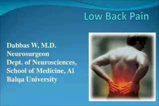
Low back pain. Presented by Mostafa kamal Abdellatif Lecturer of Anesthesia department Ain Shams University. What is back pain?. Pain that occurs in an area with boundaries between the lowest rib and the crease of the buttocks Types of back pain Acute back pain

Low back pain
E N D
Presentation Transcript
Low back pain Presented by Mostafa kamal Abdellatif Lecturer of Anesthesia department Ain Shams University
What is back pain? Pain that occurs in an area with boundaries between the lowest rib and the crease of the buttocks Types of back pain Acute back pain Pain with acut onset and rapid progressive course. Chronic back pain Pain that persists longer than the expected time period for healing greater than 3 months.
Epidemiology Low back pain that lasts at least one day and limits activity is a very common and widespread complaint. Over a lifetime, 80% of people have lower back pain,with the difficulty most often beginning between 20 and 40 years old. It is most common among women, and among people aged 40–80 years, with the overall number of individuals affected expected to increase as the population ages. Women may be more prone to raise the complaint due to pain related to osteoporosis, menstruation or pregnancy.
Classification There are many different methods for classification: 1) According to origin: Mechanical and Non mechanical 2) According to signs and symptoms: Non specific, Radicular and Specific. 3) According to chronicity: Acute, Subacute and chronic.
Etiology of back pain Acute back pain Acute low back pain is most often caused by a sudden injury to the muscles and ligaments supporting the back. It lastsfor six weeks Subacute back pain It is from six to tweleve weeks Chronic back pain : It prersists more than tweleve weeks Mechanical Endocrinologic Rheumatologic Hematologic Infection Neurologic. Referred Neoplastic Psychiatric Miscellaneous
Mechanical 85% of all low back pain Muscle, ligament, tendon strain. Discogenic disorders including herniated disc. Apophyseal joint arthritis. Spinal stenosis. Spondylolysis, spondylolisthesis. Scoliosis.
Sources of low back pain Superficial somatic - skin Deep somatic - muscle, joint, tendon, bursa, fascia Radicular - nerve root Visceral referred - sympathetic afferents Neurogenic - mixed motor sensory nerves Psychogenic - cerebral cortex
Pain intensity Minimal - mentioned in passing, normal function Mild - component of symptoms, mild dysfunction Moderate - major component of symptoms, alters function Severe - the disease symptom, incapacitating function
Diagnosis of low back pain 1) History Red flags as cancer, cauda equina syndrome, fracture and infection. 2) Examination Inspection: No obvious deformities, No erythema or Skin lesions (Zoster) Palpation: Soft Tissue: we examine 3 clinical zones (Paraspinal muscles, Gluteal muscles, Sciatic area) Bones Primarily palpating spinous processes and facets Range of movement:Flexion - 80º, Extension - 35º, Side bending - 40º each side Twisting - 3-18º Strength Neurovascular Special Tests 3) Radiology
Special tests Straigth leg raising test Raising of stretched limb in supine position, positive test detected by pain in the limb. Sciatic stretch test In case of positive SLR, lowering the stretched leg 10 degrees detect pain in foot.
FADIR test: Flexion. Adduction Internal rotation FABER test: Flexion Abduction External rotation
Hoover test Helps to determine whether pt is malingering, Should be performed in conjunction with SLR When pt is genuinely attempting to raise leg, he exerts pressure on opposite calcaneus to gain leverage
Neurological Testing Sensation Strength Reflexes
Choice to do imaging based on: Trauma, chronic steroid use = XRay Suspect abscess, cauda equina = MRI Examination New/severe sensory or strength loss = consider MRI
Treatment of back pain Mild to Moderatesingle drug v. placebo (active comparator). Moderate to Severe stable multidrug regimen - flare with withdrawal. Bed rest for 1-2 weeks Multimodality Back exercises - flexion and/or extension Aerobic exercise Medications Counterirritant topical therapies Stress management
Medications NSAIDs Muscle relaxants Analgesics Antidepressants Anticonvulsants Alpha-2 adrenergic agonists Miscellaneous
NSAIDS Improve pain vs. placebo in controlled trial Short half-life acute exacerbations, quick onset Long half-life Sustained effect Cox - 2 inhibitors Muscle relaxants Most beneficial in the first week, Shown effective in trials. Similar efficacy,Work best when combined w/ NSAIDs. Cyclobenzaprine Orphenadrine Metaxolone Chlorzoxazone Methocarbamol
Analgesics Nonnarcotic Acetaminophen Tramadol Narcotic Short acting Long acting
Antidepressents Tricyclic and tetracyclic antidepressents as. amititriptylline, maprotiline. Anticonvulsants Gabapentin Pregabalin
Interventional therapy Simple injections of plain local anaesthetic without adrenalin or cortisone. Specific management includes medial branch and sacroiliac joint blocks, and radiofrequency neurotomy. Patients with long term pain may be referred to a psychologist for cognitive behavioural therapy.