Download
1 / 37
420 likes | 622 Vues
Pleural Effusion. PLEURA. Visceral pleura envelope all surfaces of. Parietal pleura , and ribs. Hilum where pulmonary vessels, bronchi, and nerves enter the lung tissue, the parietal pleura is continuous with the visceral pleura. PLEURAL EFFUSION Normally the pleural space contains :

E N D
Visceral pleura envelope all surfaces of. Parietal pleura , and ribs. Hilum where pulmonary vessels, bronchi, and nerves enter the lung tissue, the parietal pleura is continuous with the visceral pleura.
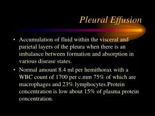
PLEURAL EFFUSION • Normally the pleural space contains: • 3.5 to 7.0 ml of clear liquid • low protein content • small number of mononuclear cells • Pleural effusion: presence of large amount of fluid in the pleural space irrespective of the underlying causes
PLEURAL FLUID FORMATION AND ABSORTION • The rate of fluid formation is 0.02 ml/kg/hour. • The rate of fluid clearance is 0.2 ml/kg/hour.
Starling’s Law Fluid movement Starling’s law: L . A [ (PCAP – PPl) – (CAP – Pl) ] L: Filtration coefficient A: Surface area Cap: Capillary Pl: Pleural
Pleural Fluid Formation and Absorption Pleural Space Intercostal Micro vessels Bronchial Micro vessels VEIN VEIN ARTERY ARTERY ? LYMPHATICS TO MEDIASTINAL NODES PLEURAL FLUID STOMA PLEURAL SPACE PARIETAL PLEURAL VISCERAL PLEURAL
Development of Pleural Effusion pulmonary capillary pressure (CHF) capillary permeability (Pneumonia) intrapleural pressure (atelectasis) plasma oncotic pressure (hypoalbuminemia) pleural membrane permeability (malignancy) lymphatic obstruction (malignancy) diaphragmatic defect (hepatic hydrothorax) thoracic duct rupture (chylothorax)
Other causes of pleural effusion: nephrotic syndrome, TB, collagen vascular disease, urinothorax, SVC syndrome, Meigs syndrome, rheumatoid arthritis, pancreatitis, yellow-nail syndrome, drugs
Symptoms symptom key symptom shortness of breath Fluid filling the pleural space makes it hard for the lungs to fully expand, causing the patient to take many breaths so as to get enough oxygen. If parietal pleura is irritated mild painor a sharp stabbing pleuritic type of pain. Some patients will have a dry cough.
Symptoms Occasionally ------> no symptoms at all. • This is more likely when the effusion collects gradually • Chest examination will reveal stony dullness, and decrease/absent breath sounds
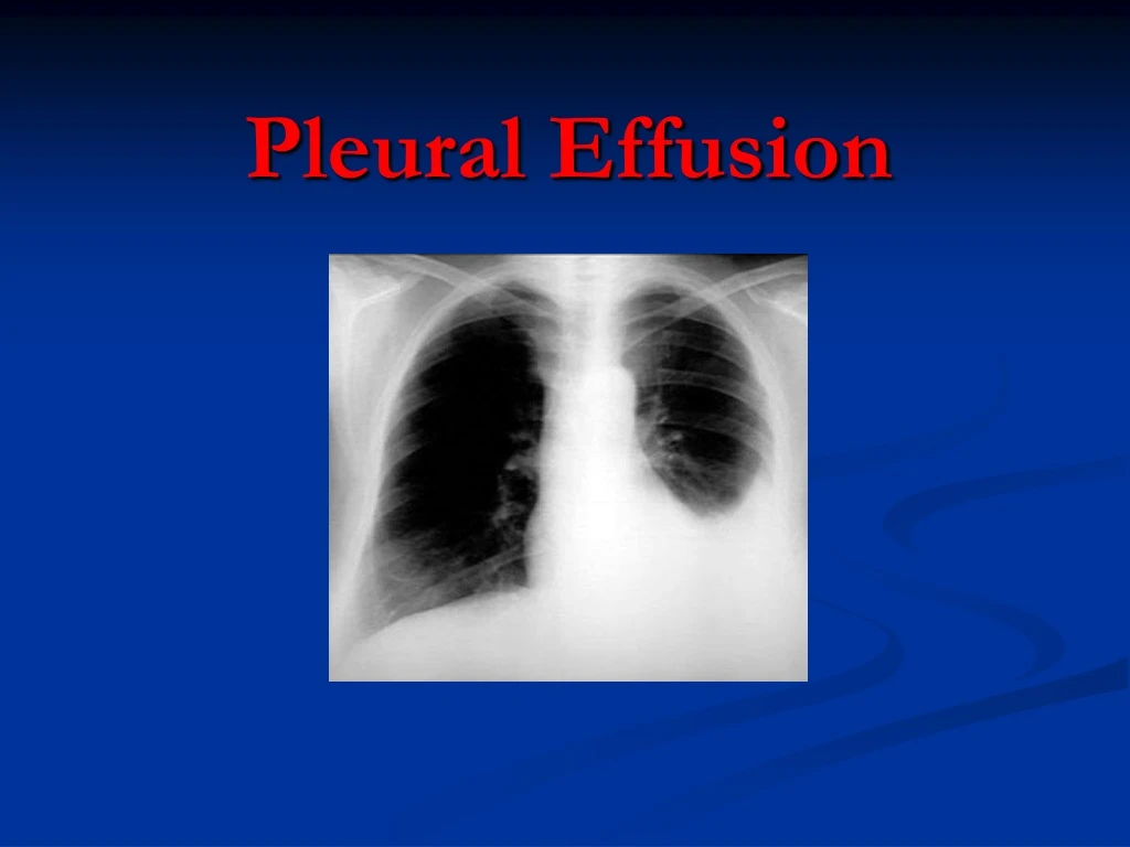
Pleural Effusion • Pleural effusion is an abnormal accumulation of fluid in the pleural space. The 5 major types of pleural effusion are: • Transudate, • Exudate, • Empyema, • Hemorrhagic pleural effusion or hemothorax and • Chylous or chyliform effusion.
History: dyspnea pleuritic chest pain cough fever hemoptysis wt. loss trauma hx cancer cardiac surgery Physical: Dullness to percussion Decreased breath sounds Absent tactile fremitus Evaluation
Causes of Pleural Effusion Other causes of pleural effusion: nephrotic syndrome, TB, collagen vascular disease, urinothorax, SVC syndrome, Meigs syndrome, rheumatoid arthritis, pancreatitis, yellow-nail syndrome, drugs Light. NEJM 2002; 346:1971 Annual incidence in the US
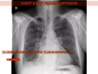
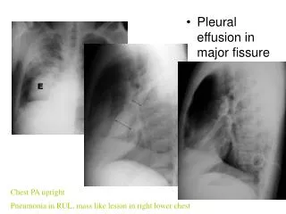
Chest X-Ray • Lateral decubitus • PA
Indications for thoracentesis • Effusions larger than 1cm height of unknown origin • No need for thoracentesis for patient with obvious cause (CHF with bilateral effusions). However: • In heart failure: febrile/pleuritic pain, unilateral, no cardiomegaly, no response to diuresis
Pleural fluid analysis Appearance Bloody Hct <1% not significant, 1-20%= CA, PE, Trauma >50% serum Hct = hemothorax Cloudy trig level >110mg/dl = chylothorax Putrid odor stain and culture = infection? Transudate vs Exudate?
Exudate v Transudate Patient’s serum protein is normal Pleural protein is less than 25 g/l =Transudate Pleural Protein more than35 g/l.= Exudate If not, Light’s criteria
Light’s Criteria Pleural fluid is exudate if one or more: Pleural fluid protein:serum protein > 0.5 Pleural fluid LDH:serum LDH > 0.6 Pleural fluid LDH > 2/3 upper limit nl serum LDH • Exudate • Pneumonia • Malignancy • Pulmonary Embolism • Transudate • CHF • Cirrhosis • Nephrotic syndrome
Exudative Effusion • Cell count - Neutrophil predom acute pleural process (pneumonia, PE) - Lymphocytic predom chronic process (Cancer, TB, CABG) • Culture/stain- infected fluid • Glucose- low level (<60mg/dl)(pneumonia, CA) • Cytology- malignancy (non-dx- thoracoscopy) • pH- parapneumonic <7.2 -must drain fluid malignant < 7.2 –poor prognosis
Lymphocytic (> 50%) CA (30-35%) TB (15-20%) Sarcoidosis PMNs Empyema Parapneumonic Rheumatoid Pulmonary infarction PMN or Lymphocytic PE Conn tissue disease Post-cardiac injury Eosinophilic (> 10%) Trauma PTX CA Asbestos, parasites Pneumonia RBC > 100,000/mm CA Trauma Pulmonary infarction EXUDATIVE EFFUSIONS
Suspected TB Adenosine deaminase (> 50 IU/L) B2 - microglobulin Lysozyme III (> 20mcg/mL) PCR (Sens 100%, Spec 95%) AFB (smear 10-20%; cx 25-50%) PPD Suspected Rheumatoid Pleural RF Low glucose Suspected SLE Serum Complement Pleural ANA LE cells prep? Suspected Pneumonia pH Suspected Pancreatitis Pleural Amylase EXUDATIVE EFFUSIONSOther Tests
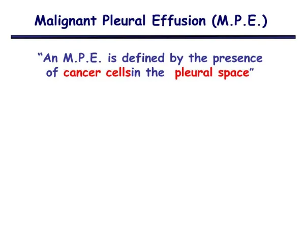
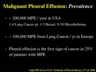
Malignant Effusions • Clinical features suggestive of malignacy: Symptoms> 1mo, absence of fever, blood-tinged fluid, chest CT suggesting malignancy • Lung >breast > lymphoma/leukemia • metastatic adenocarcinoma positive cytology 70% • Lymphoma 25-50% • Mesothelioma 10% • Squamous Cell Carcinoma 20% • Sarcoma within pleura 25% • Pleural fluid: bloody, lymphocytic, decreased or nl glucose and pH, cytology
Treatment • Thoracentesis – then treat underlying disease • Uncomplicated pneumonia – antibiotics • Hemithorax involved/empyema – tube thoracostomy +/- VATS • Malignant effusion- chest tube +/- pleurodesis (sclerosants) VATS
UNDIAGNOSED PLEURAL EFFUSIONS • 15-20% of effusions • Careful review of history, PE, meds, risk factors • Consider occult abdominal process • Consider PE
UNDIAGNOSED PLEURAL EFFUSIONSCont’d • Risk factors for TB or malignant effusion • Weight loss > 4.5 kg (10 pounds) • Fever > 38 C • Positive PPD • Large effusion (> 1/2 hemithorax) • < 95% lymphs in pleural fluid • If ANY factor present, evaluate for TB, CA
BEYOND THORACENTESIS • Pleural Biopsy • Most helpful in evaluating for TB • Limited utility for CA • Thoracoscopy • Most helpful in evaluating for malignancy
Treatment • Thoracentesis – then treat underlying disease • Uncomplicated pneumonia – antibiotics • Hemithorax involved/empyema – tube thoracostomy +/- VATS • Malignant effusion- chest tube +/- pleurodesis (sclerosants) VATS
Indications for Chest Tube • Empyema • Complicated parapneumonic effusion PH <7.2 • Hemothorax • Malignant effusion- chest tube +/- pleurodesis (sclerosants)
Pleural Biopsy • Most helpful in evaluating for TB • Limited utility for CA