Enhancing Accuracy in In Vivo Dosimetry for Radiotherapy
350 likes | 775 Vues
In vivo dosimetry is crucial for accurate radiation dose delivery in radiotherapy, ensuring patients receive the prescribed dose. This process involves measuring the dose delivered to patients directly, accounting for individual variations like patient contour, organ motion, and treatment setup. Reliable dosimeters, such as semiconductors, TLDs, and EPIDs, are essential for precise readings. Calibration and correction factors, including temperature and beam energy, must be carefully managed to maintain accuracy. Novel techniques like dose-guided radiotherapy aim to improve precision and patient outcomes.

Enhancing Accuracy in In Vivo Dosimetry for Radiotherapy
E N D
Presentation Transcript
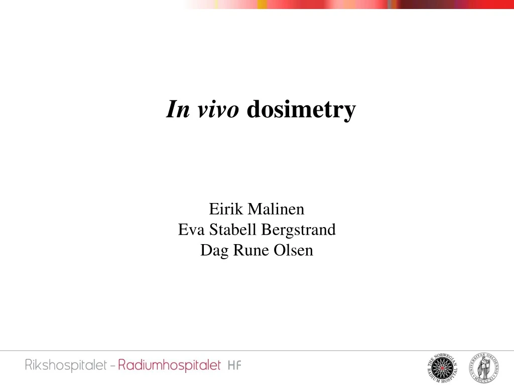
In vivo dosimetry Eirik Malinen Eva Stabell Bergstrand Dag Rune Olsen
error Probalility Prescribed dose In vivo dosimetry • In vivo: In the living • Dosimetry: Estimates of radiation dose by theory and measurement • Verification of delivered • dose to individual patients • Radiotherapy requires • accurate dose delivery
Errors in patient dose • Patient contour / planning basis (CT images) • Patient motion • Organ motion • Dose calculations (inhomogeneities, scatter) • Patient positioning • Transfer of treatment data from simulator to linac • Linac settings (energy, monitor units, field size) and calibration • Beam modifiers (blocks, wedges)
Dose measurements Point detector beam Entrance dose: wedge 2D detector array Output, SSD Wedge, curvature Patient curvature Exit dose: Thickness, density
High accuracy Low precision Low accuracy High precision Desired in vivo dosimeter characteristics • Accurate and precise • Multiple readouts • Reusability • No cables • Non-destructive readout
In vivo dosimetry principles • Point detector: • Semiconductors (diodes) • Thermoluminescent crystals • EPR (electron paramagnetic resonance) sensitive materials • …. • 2D detector, (electronic) portal imaging device; EPID: • Film • Arrays (ion chambers, semiconductors)
Dosimeter reading → absorbed dose • Absorbed dose, D: • R: dosimeter reading • ND: calibration factor • Ci: correction factor
Calibration • Under reference conditions: beam dosimeter Rcal dmax ion chamber Dcal water phantom
Example – diodes spherical droplet
Correction factors • Dosimeter reading may depend on: • Temperature • (Accumulated) Dose • Dose rate • Beam energy • Field size • ... • Accuracy may be reduced if dependence is not corrected
Temperature and sensitivity, diodes Detector temperature after placing on patient Sensitivity dependence
Accumulated dose and sensitivity, diodes • Regular calibration must be performed
Field size and sensitivity, diodes 8 or 18 MV photons Entrance (in) or exit (out)
Action level • Relative dose difference: • At what dose difference level should the treatment be revised? 1% ? 2.5 % ? 5 %? • Depends on: • dosimetric accuracy and precision • non-systematic errors • …
Methods Portal image profile
Measured dose / prescribed dose Action level: 2.5% measured dose dose after correction
Treatment planning algorithm Portal image Collapsed cone algorithm Location of normalization point 2D dose maps
Novel methods – ”dose guided radiotherapy” prescribed isodose target Backprojection of filtered dose image into patient image →OK →correction dose image
Corrections Novel methods – ”dose guided radiotherapy” bladder prostate rectum