Application of Hip Arthroscopy
970 likes | 2.38k Vues
Application of Hip Arthroscopy. Nadhaporn Saengpetch, MD. Objectives. To understand the spectrum of disease that is compatibly treated with hip arthroscopy Have a basic understanding of the relevant anatomy , history and examination for hip pain

Application of Hip Arthroscopy
E N D
Presentation Transcript
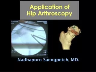
Application of Hip Arthroscopy Nadhaporn Saengpetch, MD.
Objectives • To understand the spectrum of disease that is compatibly treated with hip arthroscopy • Have a basic understanding of the relevant anatomy , history and examination for hip pain • To introduce the surgical technique and its limitation
Once upon a time…. • 1802 Dr. Phillipp Bozzini “Lichtleiter” • 1931 Dr. Micheal S. Burman 20 cadaveric hip joints
First Clinical Application : 1939 Dr. Kenji Takagi 2 Charcot joints 1 Tbc arthritis 1 Supparative arthritis J Jpn Orthop Assoc 1939
Tip of Physical Exams • Differential diagnosis to intra/extra-articular pain, pubic pain • One joint above and below • Gait : LLD, pelvic obliquity • foot-progression angle & muscle contraction
Tip of Physical Exams • Intra-articular lesion : log rolling, McCarthy hip extension sign • SI problem : FABER test • Hip flexion contracture : Thomas test • Piriformis syndrome : sit & active ER • Hip dysplasia : anterior apprehension test ( extend & ER)
Differential Diagnosis of Groin Pain 4 zones of groin pain
Labral Tears • Traumatic tears posterior hip dislocation pain/catching after twisting or slipping repetitive hyperflexion
Labral Tears N. America : 436 hips, 96% were anterior lesion (twist, pivot) McCarthy JC. JBJS Am May 2005 Asian hips were most postero-superior lesion (hyperflex, squat) Ikeda T. JBJS Br June 1988
Labral Tears • Degenerative tears OA hip relieve mechanical symptom in some pts did scope in early OA pts worsen outcome (Walton NP. Int Orthop June 2004)
Arthroscopic Classification of Hip Labral Tears *Radial flap Radial fibrillated Lage LA. Arthroscopy Dec 1996. Longitudinal Complex
Labral Tears • Hip dysplasia • selected patient • literatures devoid of studies this patient population, open acetabular osteotomy remains reasonable & well-described treatment • shollow acetabulum subluxate & distribute abnormal stress from a head on the labrum
Chondral Lesions • Lateral impact mechanism ( by GT) • Associated labral tears 55.3% (McCarthy JC. Clin Orthop 2001) • Cartilage stimulation • ACI • Future : more predictable cartilage-resurfacing procedure
Labral Lesion with Chondral Lesion • Subchondral cyst • formation • Synovial fluid • burrows beneath • the delaminating • cartilage and • subchondral bone
Risk Factors of 2º OA from Labral Tears • With developmental dysplasia • Tears > 5 years old • Full-thickness chondral lesion
Ligamentum Teres Rupture • Deep anterior groin pain • Mechanical symptoms • History of significant trauma • Associated pathology : labrum, LB, chondral damage • ? Incidence (Byrd JWT. Arthroscopy April 2004)
Snapping Hip(Coxa Sultans Interna) Iliopsoas bursitis
Synovial Abnormalities • Chondromatosis • Crystalline disease • RA/SLE • Ehler-Danlos : capsular shrinkage
Femoral Acetabular Impingement (FAI) • Leads to OA hip • anterior head-neck offset or acetabular overcoverage
Radiographic Workup • AP view • Lateral view (Cross-table) • Lateral view (Dunn, false-profile) Alpha angle Control 42º FAI pt 74 º (Notzli HP. JBJS Br March 2002)
Cam Type • Caused by shear forces of the non-spherical position of the head against the acetabulum • Anterosuperior cartilage • Predisposing factors : SCFE, abnormal epiphyseal extension, malunion neck/head fracture, and femoral retroversion
Pincer Type • Repetitive stresses of a normal neck against an abnormal acetabular rim (over-coverage) • Antero-superior labrum “coup” • Postero-inferior head “contre-coup” • Predisposing factors : acetabular protrusio/retroversion, malunion acetabulum, 2˚ from osteotomy
Pincer Type Normal Cross-over sign
Mixed Type • Combine head/cup lesions • Less isolated type (Cam 9%, Pincer 5%) (Beck M. JBJS Br Jan 2005)