Download
1 / 30

E N D
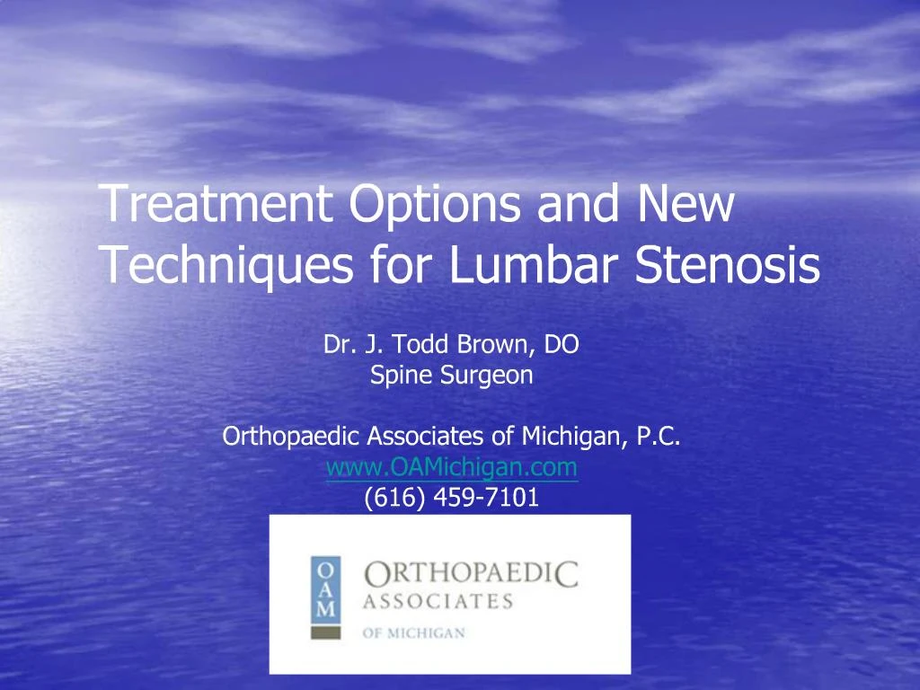
1. Treatment Options and New Techniques for Lumbar Stenosis Dr. J. Todd Brown, DO
Spine Surgeon
Orthopaedic Associates of Michigan, P.C.
www.OAMichigan.com
(616) 459-7101
2. What we�ll cover today� Basic Spine Anatomy
Spinal Stenosis
What is it�
What causes it�
Symptoms
How is it diagnosed
Treatment Options
Nonsurgical options
Surgical options
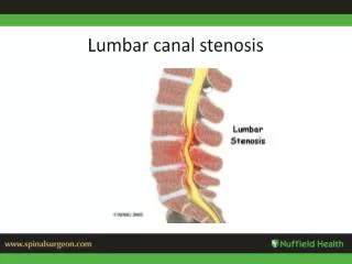
3. Your Spine Your spine consists of three primary sections:
Cervical- Neck
Thoracic- Upper back
Lumbar- Lower back
4. Spinal Anatomy
5. Spinal Anatomy
6. Spinal Anatomy
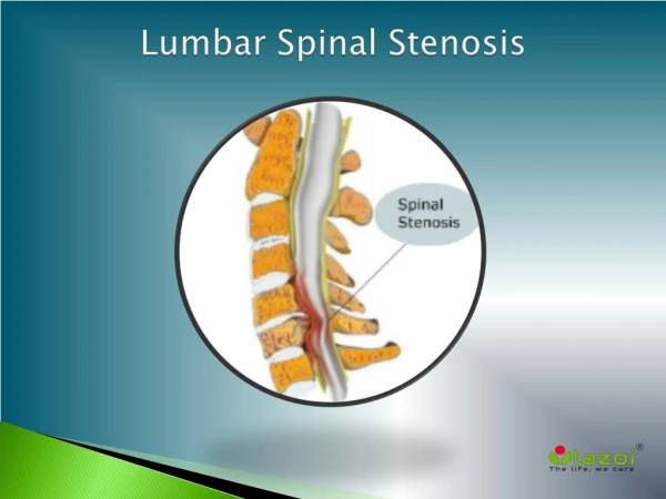
7. What is Spinal Stenosis? Narrowing of the spinal canal that places pressure on the spinal cord, cauda equina, or nerves that extend to the legs.
8. Spinal Stenosis Anatomy
9. What Causes Spinal Stenosis? Canal Narrowing caused by:
Disc bulge/herniation
Hypertrophied (thickened) Ligamentum flavum
Hypertrophied facet joints (thickened joint due to arthritis)
Spondylosis (bone spurs)
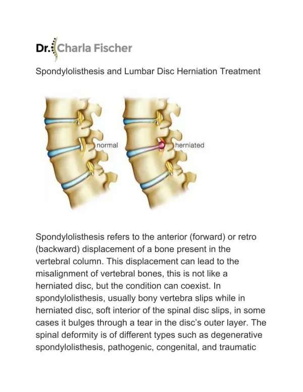
10. What Causes Spinal Stenosis? Spondylolisthesis (one bone slipping forward over another)
11. What Causes Spinal Stenosis?
12. Symptoms of Spinal Stenosis Low back pain
Pain and weakness in the legs that limits prolonged standing or walking
Numbness or tingling that radiates down the legs which tends to worsen with standing and walking
Symptoms tend to slowly worsen over time
13. Symptoms of Spinal Stenosis
Symptoms can radiate down one leg, sometimes referred to as sciatica
14. Symptoms of Spinal Stenosis If the stenosis is severe sometimes both legs can have symptoms
The location of symptoms can vary depending on which nerves are involved
15. Symptoms of Spinal Stenosis
16. How is Spinal Stenosis Diagnosed? X-rays may be recommended to evaluate the bony structure and may identify osteophytes (bone spurs) or spondylolisthesis (slipped vertebrae)
Bending x-rays may help identify potential instability
17. How is Spinal Stenosis Diagnosed? Magnetic Resonance Imaging (MRI) provides exceptional detail of bony and soft tissue anatomy
Noninvasive
No radiation
18. How is Spinal Stenosis Diagnosed? Myelography may also be used to give better definition of nerve structures
Requires direct injection of contrast into spine around nerves
A series of x-rays are then completed
E:\Spine pics\csf-fig1.jpg
19. How is Spinal Stenosis Diagnosed? Computed Tomography scan (CT or CAT scan) utilizes a computer to reconstruct x-rays into different viewing angles
Produces detailed pictures of bone and metal
Myelography may also be used with CT to give better definition of nerve structures
Requires direct injection of contrast into spine around nerves E:\Spine pics\lumbar myelo stenosis.jpg
20. Nonsurgical Treatment Options After evaluation, a doctor may recommend a variety of nonsurgical options to alleviate your pain
This may include
Physical therapy
Pain medication
Anti-inflammatories
Chiropractic care
Bracing
Activity modification
Weight loss
21. Nonsurgical Treatment Options
Epidural steroid injections may be recommended and can provide significant relief
Amount and time of relief can be unpredictable
22. Nonsurgical Treatment Options
Epidural steroid injection being performed under
x-ray guidance
23. Surgical Treatment Options Surgical Decompression
Laminectomy � removal of the lamina
Requires hospital stay
Typical recovery of 4 to 6 weeks
May require fusion with instrumentation (screws) which may lengthen hospital stay and recovery
Hemi laminectomy
Similar to laminectomy but removes less bone (hemi=half )
Potentially done on outpatient basis
Quicker recovery
Requires manipulation of nerves
E:\Spine pics\hdg7_laminectomy.jpg
24. Surgical Treatment Options
Posterior view of 2 level laminectomy
25. Surgical Treatment Options
X-ray showing evidence of hemilaminectomy
26. Surgical Treatment Options Laminectomy with instrumented fusion
May be required if stability is a concern
Hospital stay longer
Higher risks
Longer recovery with restrictions, such as a brace
27. Surgical Treatment Options AP (Front/Back view) and Lateral (Side view)
of 2 level instrumented spinal fusion
28. Surgical Treatment Options Minimally invasive techniques
X-Stop Implant
Implant device placed between spinous processes
Mimics bending or sitting
Outpatient surgery done in less than one hour
Quicker recovery
No manipulation of nerves or entrance into spinal canal
Less potential risks
Used only in select patients
Symptoms must be relieved with sitting or bending forward
29. Surgical Treatment Options
Close-up view of
X-Stop spinal implant
30. Treatment Options Which treatment is right for me?
A treatment plan can only be developed in close consultation with a spine specialist since no two problems are the same
Generally, invasive procedures are avoided unless there is:
Persistent severe symptoms
Progressive weakness or neurologic deficit
Failure of noninvasive treatment
31. Questions? Thank you for your time and attendance.
Dr. J. Todd Brown, DO
Spine Surgeon
Orthopaedic Associates of Michigan, P.C.
www.OAMichigan.com
(616) 459-7101