HERNIA dr sigid djuniawan
1.01k likes | 1.13k Vues
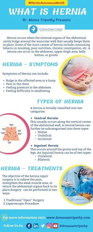
Learn about hernias, including causes, symptoms, anatomy, and treatment options to prevent complications like strangulation. Find out about different types of hernias and when surgery is necessary.

HERNIA dr sigid djuniawan
E N D
Presentation Transcript
Introduction • Protrusion of the peritoneum or preperitoneal fat through an abnormal opening in the abdominal wall • Presents as a bulge • Peritoneal contents may be trapped in “sac” • Asymptomatic bulge most common • Symptoms • Physical effects of sac and contents on surrounding tissues • Obstruction and/or strangulation of hernia sac contents
Epidemiology • 700,000 hernia repairs year • Inguinal hernias -75% of all hernias • 2/3 Indirect, remainder are direct • Incisional hernias – 15 to 20% • Umbilical and epigastric – 10% • Femoral – 5%
Epidemiology • Prevelance of hernias increases with age • Most serious complication – strangulation • 1 to 3% of groin hernias • Femoral – highest rate ofcomplications 15% to 20% • recommended all be repaired at time of discovery
Areas of Natural Weakness Used with permission from the American College of Surgeons
Anatomy • Inguinal ligament (Poupart’s) – inferior edge of external oblique • Lacunar ligament – triangular extension of the inguinal ligament before its insertion upon the pubic tubercle • conjoined tendon (5-10%)- Internal oblique fuses with transversus abdominis aponeurosis • Cooper’s Ligament - formed by the periosteum and fascia along the superior ramus of the pubis.
Inguinal Canal • Between deep and superficial inguinal rings • Boundaries • Superifical – external oblique aponeurosis • Superior – internal and transversus • Inferior – shelving edge of inguinal ligament and lacunar ligament • Posterior (floor) – transversalis fascia and aponeurosis of transversus abdominis muscle
Components of Hesselbach’s triangle include which of the following anatomic landmarks? • Pectineal ligament • Lateral border of the rectus sheath • Cooper’s ligament • Inguinal ligament • Inferior epigastric vessels
Hernia Diathesis • Varies with age • Pediatric: congenital remnant • Adult • Tissue weakness • Burst strength < abdominal wall tension • Varies with gender
Hernia Diathesis • Pediatric: major risk is premature birth • Adult • Obesity • Previous abdominal surgery • Pregnancy • Abrupt abdominal wall exertion
What is a Hernia composed of? Sac: a folding of peritoneum consisting of a mouth, neck, body and fundus. Body: which varies in size and is not necessarily occupied. Coverings: derived from layers of the abdominal wall. Contents: which could be anything from the omentum, intestines, ovary or urinary bladder.
A sliding inguinal hernia on the left side is likely to involve which of the following? • Jejunum composing the posterior wall of the sac • Ovary and fallopian tube in a female infant • Omentum • Sigmoid colon composing the posterior wall of the sac • Cecum composing the anteromedial wall of the sac
Terminology • Pantaloon – direct and indirect components • Richter’s – contains antimesenteric portion of small bowel • Sliding – involves visceral peritoneum of an organ , i.e. bladder, ovary • Littre’s – hernia contains Meckel’s diverticulum • Petit – hernia at inferior lumbar triangle • Grynfelt – hernia at superior lumbar triangle
Clinical Evaluation: History • Demographics • Age • Gender • Presentation of bulge • When, where, how • Activities that make it better or worse • Discomfort vs. pain • Signs/symptoms of bowel obstruction
Clinical Evaluation: History • Surgery: previous repairs/operations • Review of factors related to increased intra-abdominal pressure • Chronic cough • Constipation • Straining to urinate
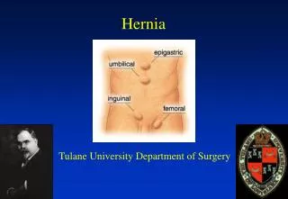
Clinical Evaluation: Location • Groin: 75% • Inguinal • Femoral • Anterior abdominal wall: 25% • Umbilical • Epigastric • Spigelian • Incisional
Hernia Pathology • Contents of hernia sac • Bowel (small and large, appendix) • Incarceration of portion of bowel wall: Richter’s hernia: Strangulation occurs without obstruction • Omentum, bladder, ovary, fallopian tubes • Sac wall may be formed by large bowel, bladder, or the ovary/tube: Sliding hernia
Hernia Pathology • Fascial defect may exist without peritoneal hernia sac • Preperitoneal abdominal wall contents may protrude through fascial defect • Preperitoneal fat • Lymph node
Hernia Pathology • Incarceration: contents of hernia sac not reducible into peritoneal cavity • Acute: fascial margins trap contents • Chronic: contents adhesed in sac • Strangulation: incarceration with compromise of blood supply • Narrow neck at greatest risk: indirect inguinal, femoral, and umbilical
Hernia Repair Indications • Asymptomatic • prevent visceral incarceration and/or strangulation • Symptomatic, non-obstructed • Treat discomfort from bulge • Prevent incarceration/strangulation • Visceral obstruction/strangulation • Release obstruction/manage viscera • Prevent recurrence
Groin Hernia • Men : Women 25 : 1 • Right : Left 2 : 1 • Femoral • Women > Men • Strangulation risk > inguinal • Inguinal • Indirect : Direct 2 : 1 • Most common in men and women
Groin Hernia • Inguinal: relationship of sac to inguinal canal determines external bulge • Movement from internal ring to scrotum • Bilateral hernias: direct 4x indirect • Indirect vs. direct hernia is intraoperative diagnosis, not clinical diagnosis • Femoral: relationship of sac to inguinal ligament determines external bulge
Groin Hernia: Inguinal • Adults • Weakness of transversalis fascia • Indirect: sac is lateral to inferior epigastric vessels • Direct: sac is medial to inferior epigastric vessels • Pantaloon: both indirect and direct • Pediatric: patent processus vaginalis
Inguinal hernia Femaleinguinal hernia Male inguinal hernia
Groin Hernia: Differential Diagnosis • Tendonitis • Muscle tear • Lymph node • Lipoma • Varicose vein • Hydrocele • Epididymitis • Spermatocele
Groin Hernia Management • Most hernias: ambulatory OR • Local/regional/general anesthesia • Prohibitive operative risk: truss
Groin Hernia Management • Acute incarceration • Reduction (taxis) • Distal traction and gentle milking • Caution: reduction en masse • Successful reduction shows visually • Urgent elective repair if reduced
Groin Hernia Management • Emergent repair • Irreducible acute incarceration • Strangulation • Fluid, electrolyte resuscitation
Groin Hernia Surgical Classification (Nyhus) • I: Indirect hernia w/normal internal ring • 2: Indirect hernia w/enlarged internal ring • 3a: Direct inguinal hernia • 3b: Indirect hernia with weak floor • 3c: Femoral hernia • 4: All recurrent hernias
Direct Inguinal Hernia • Medial to the inferior epigastric artery and vein, and within Hesselbach's triangle • acquired weakness in the inguinal floor
Indirect Inguinal Hernia • Accepted hypothesis: incomplete or defective obliteration of the processus vaginalis during the fetal period • remnant layer of peritoneum forms a sac at the internal ring • more frequently on the right
Femoral • More common in females • Up to 40% present as emergencies with hernia incarceration or strangulation • Passes medial to the femoral vessels and nerve in the femoral canal through the empty space • Inguinal ligament forms the superior border
Groin Hernia Surgery: Open • Indirect sac: high ligation • Men: ligation at internal ring • Women: ligation/excision of round ligament with closure of internal ring • Cord lipoma: excision
Operative • Bassini • Shouldice • McVay • Lichtenstein • Preperitoneal • Laparoscopic
Bassini (early 20th Century) • Transversus abdominis to Thompson’s ligament and internal oblique musculoaponeurotic arches or conjoined tendon to the inguinal ligament • Shouldice (1930s) • Multilayer imbricated repair of the posterior wall of the inguinal canal • McVay (1948) • Edge of the transversus abdominis aponeurosis to Cooper’s ligament; incorporate Cooper’s ligament and the iliopubic tract (transition suture)
BASSINI MCVAY SHOULDICE
Lichtenstein • First pure prosthestic, tension-free repair to achieve low recurrence rates
Groin Hernia Surgery: Open • Inguinal floor: tension-free repair with mesh • Anterior plug and patch • Anterior patch • Posterior patch (Stoppa)
Groin Hernia Surgery • Open tissue repair for risk of infection (example: strangulated hernia) • Laparoscopic • Indications • Recurrent hernia • Bilateral hernias • Must be able to tolerate general anesthesia • More expensive
Groin Hernia Repair Complications • Recurrence • Tissue repair: 1.3—25% • Tension-free mesh: 0.5—5% • Greatest risk is repair of previous hernia at same location
Groin Hernia Repair Complications • Chronic groin pain: up to 30% • Numbness over base of scrotum
Groin Hernia Repair Complications • Wound • Hematoma: 1.0% • Infection: 1.3% • Seroma • Infertility • Injury to vas deferens • Ischemic orchitis is uncommon • Urinary retention