Rhesus Incompatibility
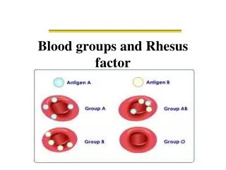
Rhesus Incompatibility. Presented by: Dr. Rozhan Yassin khalil FICOG,CABOG,HDOG,MBChB. Introduction :. Blood group is defined in two ways: 1-ABO group, allowing four different permutations of blood group ( O, A, B , AB ). 2-Rhesus system, which consists of C, D and E antigens.


Rhesus Incompatibility
E N D
Presentation Transcript
Rhesus Incompatibility Presented by: Dr. RozhanYassinkhalil FICOG,CABOG,HDOG,MBChB
Introduction: Blood group is defined in two ways: 1-ABO group, allowing four different permutations of blood group ( O, A, B , AB ). 2-Rhesus system, which consists of C, D and E antigens.
Introduction: The importance of these blood group systems is a mismatch between the fetus and mother when fetal red cells pass across to maternal circulation, as they do during pregnancy . *Sensitization of the maternal immune system to these fetal ( foreign) red blood cells may occur and subsequently give rise to the haemolytic disease of the fetus and newborn (HDFN). .
Introduction: The Rhesus system is the one most commonly associated with severe haemolytic disease.
The Inheritence of Rhesus system: The Rhesus system is coded on two adjacent genes, which sit within chromosome one . One gene codes for antigen polypeptides C/c and E/e while the other codes for the D polypeptide (Rhesus antigen). Notes that the d (little d) antigen has not been identified so it may be that women who are D negative lack the antigen altogether.
The Inheritence of Rhesus system: Antigen expression is usually dominant , where as those who have a negative phenotype are either homozygous for the recessive allele or have a deletion of that gene. In practice, only anti-D and anti-c regularly cause HDFN ( haemolytic disease of fetus and newborn and anti-D is much more common than anti-c.
The aetiology of rhesus disease: Occurrence of HDFN as a result of Rhesus isoimmunization involves three key stages : 1-First, a Rhesus negative mother must conceive a baby who has inherited the rhesus positive phenotype from the father .
The aetiology: 2-second, fetal cell must gain access to the maternal circulation in a sufficient volume to provoke a maternal antibody response . 3-finally, maternal antibody must gain transplacental access and cause immune destruction of red cells in the fetus.
Rhesus disease does not affect a first pregnancy. as the primary response is weak and consists of IgM antibodies that do not cross the placenta . there after IgG antibodies are produced and cross the placenta .
Rhesus antigen are well expressed by the fetus as early as 30 days gestation . so in a subsequent pregnancy,Whenmaternal resensetization occurs ( rhesus positive red cells pass from the baby to the maternal circulation ) IgG antibodies cross from the mother to the fetal circulation.
If these antibodies are present in sufficient quantities fetal haemolysismay occur. leading to sever anaemia that the fetus may die unless transfusion is performed.
Potential sensitizing events for Rhesus disease: 1-Miscarriage. 2-termination of pregnancy. 3-Antepartum haemorrhage. 4-Invasive prenatal testing ( chorionic villous sampling, amniocentesis and cordocentesis). 5-Delivery.
Prevalence of Rhesus disease: 1-The prevalence of D Rhesus negativity is 15% in the Caucasian population 2-It is very uncommon in orientals. 3-Rhesus disease is most common in countries where anti-D prophylaxis is not widespread, as in middle east and Russia
Prevention Rhesus iso-immunization: • _ The process of iso-immunization can be nipped in the bud by the intramuscular administration of anti-D immunoglobulins to a mother , preferably within 72 hours of exposure to fetal red cells. • Anti-D immunoglobulins mop up any circulating rhesus-positive cells before an immune response is excited in the mother.
The exact dose is determined by gestation at which sensitization occurred and size of fetomaternalhaemorrhage.- In the first trimester of pregnancy, because volume of fetal blood is so small, it is unlikely that sensitization would occur ,a standard dose of anti-D is given .
-In the second and third trimesters, : fetal blood volume is greater and because there is a possibility of a feto-maternal transfusion of several milliliters, a large dose given and kleihauer test performed
-A Kleihaure test : is a test of maternal blood to determine the proportion of fetal cells present ( relying on their ability to resist denaturation by alcohol or acid ). It will allow calculation of the amount of extra anti-D immunoglobulin required should a large transfusion have occurred. In many countries , Rhesus-negative women are given anti-D at 28 and/or 34 weeks routinely.
Fetal sign of Rhesus-Isoimmunization: • Usually clinical and ultrasound features of fetal anaemia do not usually become evident unless fetal haemoglobin is more than 5 g/dL less than the mean for gestation . • _features are not obvious unless the fetal haemoglobin is less than 6 g/dL
-fetal sign of anaemia by ultrasounds are : 1-Polyhydraminios. 2-Enlarged fetal heart. 3-Ascites and pericardial effusion. 4-Reduced fetal movements. 5-Abnormal CTG with reduced variability, eventually a ,sinusoidal, trace. 6-Hyperdynamic fetal circulation can be detected by ultrasound by measuring increased velocities in the middle cerebral artery or aorta.
Management of Rhesus disease in a sensitized women: Once a women who is D Rhesus negative has been sensitized to the D Rhesus antigen , no amount of anti-D will ever turn the clock back . -In a subsequent pregnancy , close surveillance is required .
-Rhesus disease gets worse with successive pregnancies, • so it is important to note the severity of the disease in previous pregnancies .
-Management In a sensitized women: Standard management involves monitoring antibody levels every 2 – 4 weeks from booking. Antibody levels or quantity can be described using the titer or by using IU ( international units ) as a standard quantification method.
The titer of anti-D level: Anti-D level Outcome 1. < 4 IU/mL HDFN unlikely 2. 4 – 15 IU/mL Moderate risk of HDFN 3. > 15 IU/mL High risk of hydropsfetalis
-If the antibody levels rise, the fetus should be examined for signs of anaemia. • in the past, the bilirubin concentration of amniotic fluid was determined optically to give an indirect measures of fetal haemolysis.
This involved an invasive procedure with the attendant risks of miscarriage / preterm labour and further boosting of the alloimmune response. In the last dacade , middle cerebral artery (MCA) dopplers ( peak velocity measurement)havebeen correlate reliably with fetal anaemia, the sensitivity is reported at 100% .
-A fetus with raised MCA dopplers has a high probability of anaemia. these cases are not common and the treatment should be in tertiary fetal medicine centres. Treatment options include delivery or fetal blood transfusion . delivery of the fetus is an option if the fetus is sufficiently mature.
How ever delivery of an anaemic , rapidly haemolysing premature baby is a significant risk and should not been undertaken lightly. Delivery must take place in a unit where adequate neonatal support and expertise is available and generally delivery should not be before 36-37 weeks of gestation unless there are difficulty with fetal transfusion.
* Fetal blood transfusion is life saving in a severely anaemic fetus that is too premature for delivery to be contemplated . *The aim of blood transfusion : 1.Restoring haemoglobin levels. 2.Reversing or preventing hydrops or fetal death.
*A side effect of blood transfusion it will suppress fetal erythropoesis , which reduces the concentration of antigen positive cells available for haemolysis. * The routes of blood transfusion : Depend on the gestation , the site of the cord insertion and the clinical situation.
*These routes of administration: 1.Into the umbilical vein at the point of the cord insertion . 2.into the intrahepatic vein . 3.into the peritoneal cavity ( may be the only way in low gestation). 4.into the fetal heart.
-Once a decision has been made that the fetus is severely anaemic and requires a blood transfusion, • the invasive procedure aims to first take a sample to confirm the anaemia and then infuse blood during a single puncture.
*The transfused blood should be 1.RhD negative. 2.Crossmatched with a maternal sample. 3.densely packed ( Hb around 30 g/L ),so that small volumes are used. 4.White cell depleted and irradiated. 5.screened for infection including CMV.
At delivery: -if the baby is known to be anaemic or has had multiple transfusions, a neonatologist must be present. -at delivery should exchange transfusion be required . -blood must therefore always be ready for the delivery -all babies born to Rhesus –negative womenshould have cord blood taken at delivery for a blood count, blood group and indirect coombs test.
Multipl choice: 1. The Rhesus system is coded on two adjacent genes, which sit within : • Chromosome one. • Chromosome two. • Chromosome three. • Chromosome four.
2.women who are D negative have: a.Tow little d antigen (dd). b. One capital D and one little d (Dd). c. Lack altogether antigen. d. Two capital Dantigen (DD).
3.Potential sensitizing events for Rhesus disease: a. -Miscarriage. b.-termination of pregnancy. c. trauma d.-Antepartumhaemorrhage
4.fetal sign of anaemia by ultrasounds are except: a-Enlarged fetal heart. b-Ascitesand pericardial effusion. c- increase fetal movements . d.- Abnormal CTG.
5. A transfused blood to fetus in Rhincomptibility are characterize by all except: a.RhD positive. b.Crossmatchedwith a maternal sample. c.denselypacked ( Hb around 30 g/L ). d.screenedfor infection including CMV.
Answer: • a. • C. • a,b,c,d. • C. • a.