Download
1 / 78
780 likes | 927 Vues
This document explores the intricate three-dimensional structures of immunoglobulins, detailing the antibody domain and fragment nomenclature. It discusses the structural basis of diversity in immunoglobulins, including the immunoglobulin fold, binding site categories, and the biochemical mechanisms of antigen recognition. Key concepts such as conformational changes upon binding, antibody flexibility, and the relationship between variable and constant regions are highlighted. The content elaborates on the assembly of CDRs and the canonical structures that define the antigen-antibody interactions.

E N D
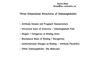
David Rose drose@oci.utoronto.ca Three-Dimensional Structures of Immunoglobulins - Antibody Domain and Fragment Nomenclature - Structural basis of Diversity - Immunoglobulin Fold - Shapes / Categories of Binding Sites - Biochemical Basis of Binding / Recognition - Conformational Changes on Binding - Antibody Flexibility - Other Immunoglobulin- like Molecules
(Fab’)2 Fv fragment
Antibody Domain and Fragment Nomenclature - Variable, Constant Regions Defined by similarity of amino-acid sequence between antibodies Each forms a structural unit (Ig fold) - Heavy Chains 1 variable domain (N-terminus): Vh 3 (IgG) or more constant domains: Ch1 - Ch3 constant domains define antibody class - Light Chains 1 variable domain (N-terminus): Vl 1 constant domain: Cl - Antigen Binding Fragment (Fab) Vl/Vh - Cl/Ch1 heterodimer - Constant (crystallizable) Fragment (Fc) Ch2-Ch3 homodimer - Variable Fragment (Fv) Vl/Vh heterodimer - Epitope: Part of the antigen recognized by the antibody - Paratope: Antibody recognition region
- Variability Plots v = num different residue types / Freq (most common) - Relationship to V(D)J gene segments
V V V V CL J J J J J CL V V J CL Light Chain V D J C Heavy Chain
Immunoglobulin Fold: Each region (V,C) forms a -sheet sandwich 110-120 residues Light Chain Variable Region
- Framework / Complementarity -Determining Regions (CDRs) (Hypervariable regions) assemble to make up the antibody binding region. - Fab/Fv binding site (combining site) 6 CDRs: 3 Light Chain + 3 Heavy Chain L1-3, H1-3 - Canonical CDR structures Chothia and Lesk L1-3, H1, H2 well defined by loop structures
Canonical forms of CDR loops. Al-lazikani, Lesk & Chothia, J Mol Biol (1997) 273:927
H3 CDR’s: 12-residue length Al-lazikani, Lesk & Chothia J Mol Biol (2000) 295:979
Shapes / Categories of Binding sites • - Flat: • mostly surface residues from both antibody and antigen. Frequently • discontinuous regions of antigen • - Groove / Crevice: • Binds to stretches of antigen, usually continuous • - Pocket: • Usually small molecule antigens, tight loops, or ends of polymers that • penetrate a small pocket.
Combining Site Shapes Pocket Groove Flat
Types of Antigens: 1. Small Molecules (haptens) Pocket-shaped shape complementarity electrostatics / hydrogen bonds (enthalpy-driven) High association constants (108 - 109 M-1)
2. Proteins a. discontinuous flat shaped hydrophobic (elimination of water) Van der Waals hydrogen bonds some entropic contribution Buried surface ~700-800 Å2 Moderate - high association constants (106 - 108 M-1)
b. continuous groove / crevice extended loop on antigen (usually -turn but can be -helix) higher entropic cost Hydrophobic, entropic, van der Waals shape complementarity Induced fit of antibody and/or antigen
Fab F11.2.32 HIV-1 protease peptide complex Lescar et al, J Mol Biol (1997) 267: 1207
Fab 17/9 complex with peptide from Influenza virus hemagglutinin Rini, Schulze-Gahmen & Wilson (1992) Science 255:959
MRK-16 Fab structure: Vasudevan, Tsuruo and Rose, J. Biol Chem (1998) 273:25413
Jean M. H. van den Elsen, Douglas A. Kuntz, Flip J. Hoedemaeker, and David R. Rose Antibody C219 recognizes an -helical epitope on P-glycoprotein PNAS 96: 13679-13684
3. Carbohydrate / polysaccharide Groove - shaped (chain binder) or pocket-shaped (end binder) Hydrophobic (especially aromatic) some hydrogen bonds water can be used as coordinating ligand High entropic cost Lower buried surface (500-600 Å2) Lower association constants (104 - 105 M-1)
Fab Se155.4 complex with Salmonella cell-surface antigen Cygler, Rose & Bundle (1991) Science 253:442
Binding of cholera O1 antigen to Fab S-20-4 Villeneuve et al, PNAS (2000) 97:8433
Conformational changes / flexibility Induced fit antibody binding site plasticity epitope Antibody flexibility domain:domain interaction changes antigen binding tether links in intact IgG structure
Antibody conformational change on complexation: Fab 17-IA with HRV-14 Smith et al (1996) Nature 383:350
Epitope Conformational change on complexation : HIV Protease
Epitope conformational changeon complexation: Flu hemagglutinin
Crystal Structure of an Intact IgG Harris LJ, Skaletsky E, McPherson A. (1998) J Mol Biol 275:861