Download
1 / 60
600 likes | 619 Vues
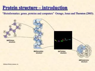
Protein informatics & structure-based analysis. Ming-Jing Hwang ( 黃明經 ) 中研院生醫所 N121 (02) 2789-9033 http://gln.ibms.sinica.edu.tw/. Protein Informatics: Objective.

E N D
Protein informatics & structure-based analysis Ming-Jing Hwang (黃明經) 中研院生醫所 N121 (02) 2789-9033 http://gln.ibms.sinica.edu.tw/
Protein Informatics: Objective • Given a protein query (sequence or structure), extract, by in silico methods, as much information/knowledge as possible about this protein.
Protein Informatics: Approaches • Homology search/knowledge transferring • Non-homology-based
Protein Informatics: contents • Primary Databases: sequence & structure • Secondary (derived) Databases: motifs, domains, families, functional sites, etc. • Tools and Webservers
NAR database Distribution of NAR Molecular Biology Database Collection Web site.
Block logos Residues that contribute to one of the blocks returned by BLOCKS database after submission of the PI3-kinase p100a sequence. (A) 4 homologues (B) 31 homologues.
Ncbi http://www.ncbi.nlm.nih.gov/Sitemap/ResourceGuide.html
Protein Data Bank (PDB) http://www.rcsb.org/pdb/home/home.do
Structure Visualization Tools • Interactive viewers KiNG (http://kinemage.biochem.duke.edu/software/king.php) Jmol (http://jmol.sourceforge.net/) WebMol (http://www.cmpharm.ucsf.edu/cgi-bin/webmol.pl) • Plugin viewers RasMol (http://www.bernstein-plus-sons.com/software/rasmol/) Swiss PDB Viewer (http://au.expasy.org/spdbv/) • Molecular graphics software Cn3D (http://www.ncbi.nlm.nih.gov/Structure/CN3D/cn3d.shtml) PyMOL (http://pymol.sourceforge.net/)
Analyzing Structure-Function Relationship Understanding Bioinformatics (Chap. 14) Marketa Zvelebil & Jeremy O. Baum
Why structure-function relationship? • Function is determined by structure (similar functions share structural fold). • Structure can provide more detailed info about function (e.g. binding sites). • With structure, many more analyses can be performed. • Reminder: however, examples exist of proteins with different functions having the same fold, and vice versa.
Str. alignment Structure-based alignment When sequence fails to reveal evolutionary kinship, structure often can.
a/b protein; parallel b sheet (b-a-b motifs) active site at C-terminal end of the barrel highly common for enzymes (>900 structures in CATH) diverse functions (divergent vs. convergent evolution) The TIM barrel fold
TIM barrel is one of heavily used functional units Gerstein & Hegyi, 1998
TIM-barrel proteins have diverse functions Non-enzyme
Similar fold but not fun. Two immunoglobulin-like b-sandwich folds with distinct functions
Different types (structures) of phosphopeptide-binding proteins SH2 (Src homology domain) PTB (phospho-tyrosine binding domain) PH (pleckstrin homology)
Domain cutting: Cbl as an example (Domain as a unit for structure comparison)
(interact with phosphotyrosine) Structural alignments reveal conserved functional sites (VAST results)
DALI finds Grb10 SH2 (DALI ignores seq order)
P53 has many interacting partners How to find binding sites? Two types: (1) large areas on surface (2) pockets or clefts
The interaction regions of survivin : using surface properties (charges, etc.) (sulfate/phosphate binding)
Binding site characteristics: patch of conserved hydrophobic or polar residues; mainly b-sheets or long loops; clusters of either hydrophobic or aromatic residues at the interface p53-ASPP2 ProMate: an automatic interface-prediction program
Partner presently unknown ASPP2 or DNA PPI-PRED predicts three PPI sites (in red) for p53.
Identification of p53 binding sites using ConSurf (PSI-BLAST + ClustalW)
Calculation of solvent-accessible surface can identify clefts or holes, which are potential binding sites. SURFNET, Pocket-Finder, and Q-SiteFinder are three programs that can do this.
DHFR Lignad binding-site programs (Q-SiteFinder and Pocket-Finder) find probable cavities where ligands can bind.