ASMP and PECAM-1 Staining in Early Metastases: Immunohistochemical Analysis
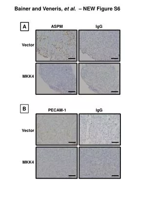
This study presents representative images from immunohistochemistry of frozen sections harvested at 14 dpi, focusing on early metastases. Panel A illustrates ASMP IgG vector staining, while Panel B showcases PECAM-1 IgG vector staining, both accompanied by associated negative controls using Rabbit IgG. These findings enhance the understanding of the expression patterns in metastatic tissues, providing critical insights into the immunological landscape during early tumor progression. Scale bars in the images represent 100 µm.

ASMP and PECAM-1 Staining in Early Metastases: Immunohistochemical Analysis
E N D
Presentation Transcript
Bainer and Veneris, et al. – NEW Figure S6 A ASPM IgG Vector MKK4 B PECAM-1 IgG Vector MKK4
Supplemental Figure SX. Immunohistochemistry of early metastases and associated negative controls.Immunohistochemistry of frozen sections from tissues harvested at 14 dpi. Panel A Representative images of ASPM staining and Panel B PECAM-1 staining are shown with associated negative controls. Negative controls were performed using Rabbit IgG as described in the Methods. Scale bars represent 100 µm.