BACK OF THE LEG
320 likes | 878 Vues
KSU College of Medicine Anatomy (121 ANA). BACK OF THE LEG. Bilal M.. K. Marwa. BACK OF THE LEG. KSU College of Medicine Anatomy (121 ANA). هذه سلايدات المحاضرة التي ألقيتها لقروب أ يوم الأثنين 25/5/2009، حيث أنهم تقبلوني بينهم، علمًا أن د/ فوهره كانت مقررة عليه المحاضرة،

BACK OF THE LEG
E N D
Presentation Transcript
KSU College of Medicine Anatomy (121 ANA) BACK OF THE LEG Bilal M.. K. Marwa
KSU College of Medicine Anatomy (121 ANA) هذه سلايدات المحاضرة التي ألقيتها لقروب أ يوم الأثنين 25/5/2009، حيث أنهم تقبلوني بينهم، علمًا أن د/ فوهره كانت مقررة عليه المحاضرة، لم أرفع هذه المحاضرة إلا لعموم 428 سواء كانوا (أ) او الشرفاء من (ب) الذين سلموا من العقد النفسية، والذين إن وجدوا بي ما لا يعجبهم قالوا لي بكل صراحة، لأننا يفتضر بنا أن نكون إخوان إن وجدتم بي ما تودون نصحي به فلا أظن أن من الصعب الوصول لي في أي وقت.. أما حركات الطفولة فقد كبرنا منها، ممن هدفهم طفولي، كاعتبار كل ما يصدر منا تنافسًا، والحقيقة أنه تعاون BACK OF THE LEG Bilal M.. K. Marwa
KSU College of Medicine Anatomy (121 ANA) BACK OF THE LEG Bilal M. K. Marwa
KSU College of Medicine Anatomy (121 ANA) • Skin • Cutaneous Nerves • - Superphicial Veins BACK OF THE LEG Bilal M.. K. Marwa
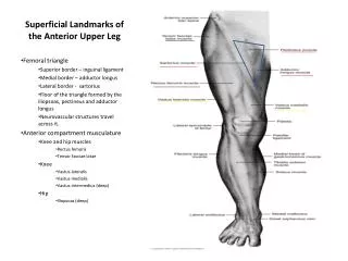
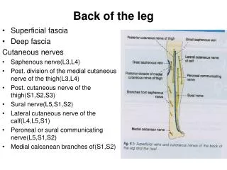
Cutaneous Nerves Skin of the back of the leg • Posterior cutaneous nerve of the thigh supplies: • skin over the poplitealfossa • Upper part of the back of the leg
Cutaneous Nerves Skin of the back of the leg • Lateral cutaneous nerve of the calf: branch of the common peroneal nerve supplies: • Skin on upper part of posterolateral surface of the leg
Cutaneous Nerves Skin of the back of the leg • Sural nerve: branch of tibial supplies • Skin on the lower part of the posterolateral surface of the leg
Cutaneous Nerves Skin of the back of the leg • Saphenous nerve: branch of femoral nerve: • Branches supply skin on posteromedial surface of the leg
Superficial veins • Small Saphenous vein: • Beginning: lateral part of dorsal venous arch of the foot. • Route: • ascends behind lateral maleoulus (with sural nerve) • Follows lateral border of tendocalcaneous then runs up the middle of back of the leg • Pierces deep fascia passing between 2 heads of gastrocnemiusinlowerpart of poplitealfossa • End: in popliteal vein (variation) • Tributaries: • Numerous small veins from back of the leg • Communicating veins with deep leg veins • Anastomosing branches running upward and medially to join greater saphenous vein
Lymph Vessels • Come from: skin and superficial fascia on the back of the leg. • Drainage route: upward, then • Either forward around medial side of leg, ending in vertical group of superficial inguinal nodes • Or Drain into popliteal nodes.
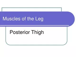
KSU College of Medicine Anatomy (121 ANA) • Muscles • Superficial group • Deep Group BACK OF THE LEG Bilal M.. K. Marwa
Contents of the Posterior Fascial Compartment of The Leg • A septum called deep transverse fascia divides the muscles of the posterior compartment into (1) superficial and (2) deep • Blood supply: posterior tibial artery • Nerve supply: Tibial nerve • 3 muscles • Gastrocnemius • Plantaris • Soleus • 4 muscles • popliteus • Flexor digitorumlongus • Flexor hallucislongus • Tibialisposterior
Superficial Group • Action: together act as powerful plantar flexors of the ankle joint • Providing main propulsive force in walking and running
KSU College of Medicine Anatomy (121 ANA) BACK OF THE LEG Bilal M.. K. Marwa
Superficial group:1. Gastrocnemius • Origin: • Lateral head: lateral condyle of the femur. • Medial head:above the medial condyle • popliteal surface of the femur • Insertion: Via tendocalcaneus into posterior surface of calcaneus • Action: plantar flexion of foot, flexion of knee
Superficial group: 2. Plantaris • Small fusiform muscle Similar to palmarislongus in forearm • May be absent or doubled • Origin: lateral supracondylar ridge of the femur. • Insertion: into posterior surface of calcaneus • long ribbon- like tendon descends between Gastrocnemius & soleus. • then on medial side of tendocalcaneus into the back of the calcaneus (they don’t merge) • Action: plantar flexion of foot, help in flexion of knee
Superficial group:3. Soleus • A broad flat muscle that forms the main bulk of the calf. • Origin: Shafts of tibia and fibula • Insertion: Via tendocalcaneus into posterior surface of calcaneus • Action: plantar flexion of foot
Deep group:1. Popliteus • Origin: Lateral surface of lateral condyle of femur • Arises intracapsular, takes a partial origin from the mensicus • Its tendon separates the mensicus from the legament of the knee, to make it freer and adapt to condylar surfaces of femur and tibia • Insertion: posterior surface of the tibia above the soleal line. • Action: flexion of the knee, • unlocking knee joint (lateral rotation of femur on tibia)
Deep group:2. FlexurDigitorumLongus • Origin: Posterior surface of shaft of tibia • Insertion: Bases of distal phalanges of lateral four toes • Each pierces the tendon of flexor digitorumbrevis of the sole • Action: • Planter flexion of the terminal phalanx of the lateral 4 toes. • Assists in planter flexion of the foot
Deep group:3. FlexurHallucisLongus • Origin: Posterior surface of shaft of fibula • Insertion: Base of distal phalanx of big toe • Action: • Planter flexion of the distal phalanx of the big toe • Assists in planter flexion of the foot. • Maintenance of medial longitudinal arch of the foot
KSU College of Medicine Anatomy (121 ANA) BACK OF THE LEG Bilal M.. K. Marwa
Deep group:4. Tibialis Posterior • Origin: • Back of interosseous membrane. • Back of tibia lateral to vertical line • Back of fibula medial to medial crest • Insertion: • All tarsus except talus. • The main insertion into tuberosity of the navicular bone. • It is also inserted into the base of 2nd,3rd& 4th metatarsal bones.
Deep group:4. Tibialis Posterior • Action: • Planter flexion of the distal phalanx of the big toe • Assists in planter flexion of the foot. • Inverts foot at subtalar and transverse tarsal joints • Supports medial longitudinal arch of the foot
KSU College of Medicine Anatomy (121 ANA) • Supply • Nerve Supply • Arterial Supply BACK OF THE LEG Bilal M.. K. Marwa
Arterial Supply:Posterior Tibial Artery • Begins as the poploteal artery divides to give posterior & anterior tibial arteries. • Begin: at level of the distal border of popliteus muscle • Route: • Passes downward deep to Gastrocnemius & soleus & deep to transverse fascia of the leg • It descends on posterior surface of tibialis posterior • Its lower part lies on back of tibia covered by skin & fascia only • It passes behind the medial malleolus to the sole deep to flexor retinaculum • Termination: divide into medial and lateral plantar aa.
Arterial Supply:Posterior Tibial Artery • Branches: • Peroneal Artery (large) • Arises close to the origin of posterior Tibial a. • Gives nutrient artery to the fibula & descends behind it. • Gives muscular branches • End: Shares in anastomosis around the ankle • Perforating branch: pierces interosseous membrane to reach lower part of front of leg • Muscular branches • Nutrient artery to tibia • Anastomotic branches
Nerve Supply:Tibial Nerve • Begin: Larger of the 2 terminal branches of sciatic nerve in the lower 1/3 of back of thigh • Route: • It bisects the poplitealfossa • It passes deep to the Gastrocnemius and soleus • It lies on posterior surface of tibialis posterior • It accompanies the posterior tibial artery. • N. medial to a., then crosses to become lateral • It passes behind the medial malleolus to reach the sole, deep to flexor retinaculum
Nerve Supply:Tibial Nerve • Branches: • Muscular branches • Medial calcaneal branch to • Articular to ankle joint • End: divide into medial and lateral plantar nn.
KSU College of Medicine Anatomy (121 ANA) Clinical Note BACK OF THE LEG Bilal M.. K. Marwa
Deep Vein Thrombosis and Long-Distance Air Travel • Passengers who sit immobile for hours on long-distance flights are very prone to deep vein thrombosis in the legs. • Thrombosis of the veins of the soleus muscle gives rise to mild pain or tightness in the calf and calf muscle tenderness. • However, deep vein thrombosis can also occur with no signs or symptoms. • Should the thrombus become dislodged, it passes rapidly to the heart and lungs, causing pulmonary embolism, which is often fatal. • Preventative measures include stretching of the legs every hour to improve the venous circulation