Download
1 / 43
480 likes | 1.09k Vues
Chapter 42. Circulation and Gas Exchange. Figure 42.3a. (a) An open circulatory system. Heart. Hemolymph in sinuses surrounding organs. Pores. Tubular heart. Figure 42.3b. (b) A closed circulatory system. Heart. Interstitial fluid. Blood. Small branch vessels in each organ. Dorsal

E N D
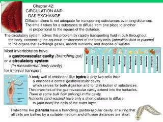
Chapter 42 Circulation and Gas Exchange
Figure 42.3a (a) An open circulatory system Heart Hemolymph in sinuses surrounding organs Pores Tubular heart
Figure 42.3b (b) A closed circulatory system Heart Interstitial fluid Blood Small branch vessels in each organ Dorsal vessel (main heart) Auxiliary hearts Ventral vessels
(a) Single circulation Figure 42.4a Gill capillaries Artery Heart: Atrium (A) Ventricle (V) Vein Body capillaries Key Oxygen-rich blood Oxygen-poor blood
(b) Double circulation Figure 42.4b Pulmonary circuit Lung capillaries A A V V Right Left Systemic capillaries Systemic circuit Key Oxygen-rich blood Oxygen-poor blood
Amphibians Figure 42.5a Pulmocutaneous circuit Lung and skin capillaries Atrium (A) Atrium (A) Right Left Ventricle (V) Key Systemic capillaries Oxygen-rich blood Oxygen-poor blood Systemic circuit
Reptiles (Except Birds) Figure 42.5b Pulmonary circuit Lung capillaries Right systemic aorta Left systemic aorta A Atrium (A) Incomplete septum V Ventricle (V) Right Left Key Systemic capillaries Oxygen-rich blood Oxygen-poor blood Systemic circuit
Mammals and Birds Figure 42.5c Pulmonary circuit Lung capillaries A Atrium (A) V Ventricle (V) Left Right Key Systemic capillaries Oxygen-rich blood Oxygen-poor blood Systemic circuit
Figure 42.5 Mammals and Birds Amphibians Reptiles (Except Birds) Pulmonary circuit Pulmonary circuit Pulmocutaneous circuit Lung capillaries Lung and skin capillaries Lung capillaries Right systemic aorta Left systemic aorta Atrium (A) Atrium (A) A A A A Incomplete septum Incomplete septum V V V V Right Left Right Left Left Right Ventricle (V) Systemic capillaries Systemic capillaries Systemic capillaries Systemic circuit Systemic circuit Systemic circuit Key Oxygen-rich blood Oxygen-poor blood
Superior vena cava Capillaries of head and forelimbs Figure 42.6 Pulmonary artery Pulmonary artery Capillaries of right lung Capillaries of left lung Aorta Pulmonary vein Pulmonary vein Left atrium Right atrium Left ventricle Right ventricle Aorta Inferior vena cava Capillaries of abdominal organs and hind limbs
Aorta Pulmonary artery Figure 42.7 Pulmonary artery Right atrium Left atrium Semilunar valve Semilunar valve Atrioventricular valve Atrioventricular valve Right ventricle Left ventricle
Atrial and ventricular diastole Figure 42.8-1 1 0.4 sec
Atrial and Atrial systole and ventricular ventricular diastole diastole 2 Figure 42.8-2 1 0.1 sec 0.4 sec
Atrial and Atrial systole and ventricular Ventricular systole and atrial ventricular diastole diastole diastole 2 Figure 42.8-3 1 0.1 sec 0.3 sec 0.4 sec 3
Figure 42.9-1 1 SA node (pacemaker) ECG
Figure 42.9-2 1 2 AV node SA node (pacemaker) ECG
Figure 42.9-3 1 2 3 AV node SA node (pacemaker) Bundle branches Heart apex ECG
Figure 42.9-4 1 2 3 4 AV node SA node (pacemaker) Bundle branches Purkinje fibers Heart apex ECG
Valve Figure 42.10a Basal lamina Endothelium Endothelium Smooth muscle Smooth muscle Connective tissue Connective tissue Capillary Artery Vein Arteriole Venule
Figure 42.13 Direction of blood flow in vein (toward heart) Valve (open) Skeletal muscle Valve (closed)
3 2 1 Figure 42.18a Collagen fibers Platelet plug Fibrin clot Platelet Fibrin clot formation Clotting factors from: Platelets Damaged cells Plasma (factors include calcium, vitamin K) Enzymatic cascade Prothrombin Thrombin Fibrinogen Fibrin
Figure 42.18b Fibrin clot Red blood cell 5 m
1 2 3 4
Figure 42.19 Stem cells (in bone marrow) Myeloid stem cells Lymphoid stem cells B cells T cells Erythrocytes Basophils Neutrophils Lymphocytes Monocytes Eosinophils Platelets
Tracheoles Muscle fiber Mitochondria Figure 42.24 2.5 m Body cell Tracheae Air sac Tracheole Air sacs Trachea Air External opening
Figure 42.22 Coelom Gills Gills Tube foot Parapodium (functions as gill) (a) Marine worm (c) Sea star (b) Crayfish
PO (mm Hg) in blood 2 Figure 42.23 O2-poor blood Gill arch O2-rich blood Lamella Blood vessels Gill arch Water flow Operculum Water flow Blood flow Countercurrent exchange PO (mm Hg) in water 2 150 120 90 60 30 Gill filaments Net diffu- sion of O2 140 110 80 50 20
O2-poor blood Figure 42.23b O2-rich blood Lamella Water flow Blood flow Countercurrent exchange PO (mm Hg) in water 2 150 120 90 60 30 Net diffu- sion of O2 140 110 80 50 20 PO (mm Hg) in blood 2
Figure 42.25a Nasal cavity Pharynx Left lung Larynx (Esophagus) Trachea Right lung Bronchus Bronchiole Diaphragm (Heart)
Branch of pulmonary vein (oxygen-rich blood) Figure 42.25b Branch of pulmonary artery (oxygen-poor blood) Terminal bronchiole Alveoli Capillaries
Figure 42.28 1 2 Air inhaled. Air exhaled. Rib cage expands. Rib cage gets smaller. Lung Diaphragm
Boyle’s Law • P=1/V
Homeostasis: Blood pH of about 7.4 Figure 42.29 CO2 level decreases. Stimulus: Rising level of CO2 in tissues lowers blood pH. Response: Rib muscles and diaphragm increase rate and depth of ventilation. Carotid arteries Aorta Sensor/control center: Cerebrospinal fluid Medulla oblongata
1 Inhaled air 8 Exhaled air Figure 42.30a 2 Alveolar spaces Alveolar epithelial cells CO2 O2 Alveolar capillaries 7 Pulmonary arteries 3 Pulmonary veins 6 4 Systemic veins Systemic arteries Heart Systemic capillaries CO2 O2 Body tissue 5 (a) The path of respiratory gases in the circulatory system
Figure 42.UN01 Iron Heme Hemoglobin
100 O2 unloaded to tissues at rest Figure 42.31a 80 O2 unloaded to tissues during exercise 60 O2 saturation of hemoglobin (%) 40 20 0 0 20 40 60 80 100 Tissues at rest Tissues during exercise Lungs PO (mm Hg) 2 (a) PO and hemoglobin dissociation at pH 7.4 2
PO (mm Hg) 2 100 Figure 42.31b pH 7.4 80 pH 7.2 Hemoglobin retains less O2 at lower pH (higher CO2 concentration) 60 O2 saturation of hemoglobin (%) 40 20 0 0 20 40 60 80 100 (b) pH and hemoglobin dissociation
Body tissue CO2 transport from tissues Figure 42.32a CO2 produced Interstitial fluid CO2 Plasma within capillary CO2 Capillary wall CO2 H2O Hemoglobin (Hb) picks up CO2 and H+. Red blood cell H2CO3 Hb Carbonic acid HCO3 Bicarbonate H+ HCO3 To lungs
To lungs Figure 42.32b CO2 transport to lungs HCO3 H+ HCO3 Hemoglobin releases CO2 and H+. Hb H2CO3 H2O CO2 CO2 CO2 CO2 Alveolar space in lung