Download
1 / 44
440 likes | 644 Vues
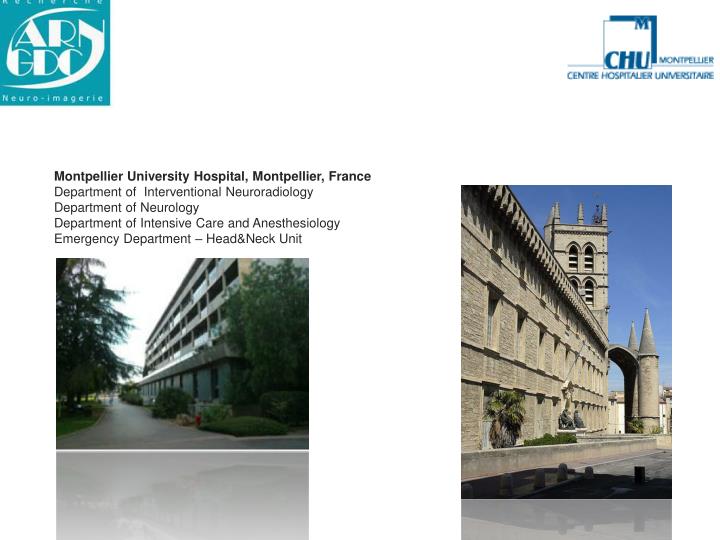
Montpellier University Hospital, Montpellier, France Department of Interventional Neuroradiology Department of Neurology Department of Intensive Care and Anesthesiology Emergency Department – Head&Neck Unit. FIBRINOLYSE IV. Intérêt rt-PA IV dans les 3H (NINDS,1995)

E N D
Montpellier University Hospital, Montpellier, France Department of Interventional Neuroradiology Department of Neurology Department of Intensive Care and Anesthesiology Emergency Department – Head&Neck Unit
FIBRINOLYSE IV • Intérêt rt-PA IV dans les 3H (NINDS,1995) mRs 0 &1:50% vs 38% (12% bénéfice absolu ou 30% bénéfice relatif) • Extension de la fenêtre thérapeutique :4H30 (ECASS3, 2008) mais avec un bénéfice relatif plus faible • Fibrinolyse IV associée à une augmentation significative du risque HIC
ECASS 3W.Hacke, N.Engl J Med,2008;259:1317 Randomisée, multicentrique,rt-PA entre 3H et 4H30 contre placebo, outcome à J90 52.4% 45.2% Efficacité marginale, bénéfice absolu du rt-PA 7.2%( p=0.04) Hemorragies IC rt-PA: 27% vs17.6% (p=0.001) NNT=17
FIBRINOLYSE IVAnalyse critique • Selon la severité de l’AVCI • Infarctus etendu :ECASS I& II • NIHSS> 20 • RISQUE HEMORRAGIQUE ACCRU • Selon le siège de l’occlusion:taux recanalisation • 8.7% ACI • 35.2% M1-ACM • 53.8% M2-ACM • 65.9% M3-ACM Del Zoppo,ann.neurol.1992;1:78-86
SCANNER C-/ AVC PHASE AIGUE Signes précoces ACM avant 3H(+75% des cas) Hypodensite NGC Effacement ruban cortical insulaire Augmentation de la teneur en H2O (hypodensité corrélée à l’infarctus final; œdème sans hypoattenuation corrélé à rVSC=tissu viable) (von Kummer, Radiology, 2001) Accord inter-observateur médiocre Score ASPECTS/ Thrombolyse IV(Dzialowski, Stroke 2006) 7 favorable; 2 risque hémorragique Signe ACM hyperdense= thrombus Sensibilité faible (27-34%), sans valeur pronostique
SELECTION DES PATIENTS SCANNER DE PERFUSION Couverture anatomique restreinte:20mm irradiation non négligeable ( 3mSev) Tracking de bolus Infarctus : rCBV cartographie (< 2.5ml/100mg) Pénombre : MTT. cartographie (>145%) Neurotherapeutics. 2011;8(1):19-27 Neuroimaging Clin N Am. 2011;21(2):215-38
P RadioGraphics 2006; 26:S75-S95 INFARCTUS PENOMBRE ↑MTT ↓CBV ↓ CBF
SELECTION DES PATIENTS IRM DWI/FLAIR MISMATCH • Circulation antérieure: • Score ASPECTS:>7 (facteur de bon pronostic) A.Demchuk, Stroke, 2005;36:2110 • Transposition cartographie ADC P.Barber ,J Neurol Neurosurg Psychiatry.2005;76:1528 K.Kimura, Stroke, 2008;39:2388 (mauvais pronostic si ≤ 5) T.Nezu, Neurology 2010 (aspects DWI≤ 5corrélé à une augmentation du taux sICH( OR 4.7); ≤ 4 corrélé au taux de morbidité(OR 3.6) • volume: DWI lesion vol facteur prédictif HIC M.Lansberg, Stroke, 2007;38:2275 (OR 1.42 pour 10ml DWI) • Circulation postérieure:pc-ASPECTS ≥8 V.Puetz, Stroke.2008;39:2485
M1 M4 C L I M5 M2 CI M3 M6 Score ASPECTS (0-10) : étendue AVCI territoire ACM. Coter 1 point = normal; Coter 0= ischémie régions corticale (M1, M2, M3, M4, M5, M6 et I) sous corticale (C, CI et L). M1: cortex antérieur ACM, M2: cortex latéral ACM, M3: cortex postérieur ACM, M4, M5 et M6 sont les points respectivement au dessus de M1, M2 et M3 ; I: cortex insulaire C: tête noyau caudé, L: noyau lentiforme et CI: genou capsule interne.
Radiology: : December 2010 Time from Symptom Onset in Acute Stroke Petkova et al
DIFFUSION/PERFUSION MISMATCH Bar.Ch,58ans,hémiplegie g masive, NHISS=20, <3H, ASPECTS =5
VOLUME INFARCTUS CEREBRAL ASPECTS =2,deficit BF gauche, 4H30
AVC Cartographie du TTP Nécrose (ADC) pénombre Parenchyme sain
Imagerie de diffusion b=10000i : phase aigue Imagerie FLAIR : 5 jours après AVC
Thrombectomie mécanique CATCH MERCI PENUMBRA SOLITAIRE
Technical considerations General Anesthesia & Femoral Approach Guiding catheter:6F for VA; 8F or 9F balloon guiding catheter for ICA (aspiration during system pull-back) Microcatheterat least .021 in of ID Microguidewire .014-.016 in Bolus of heparine IV(1000 IU after femoral puncture plus 1000 IU at the end of first hour), no antiplatelet agents Solitaire FR eV3 After the procedure: no anticoagulation therapy at least for 24 hours, CT after the procedure and CT or MRI the day after.
Protocol foracute stroke intervention Rescue (failed IV Fibrinolysis/MCA) IV Fibrinolysis 0.9 mg/kg (IV bolus 10%) Clinical Revaluation at 60 minutes If NIHSS > 7 Thrombectomy Combined/Bridging (ICA-MCA Tandem, Carotid ‘T’, BA) IV Fibrinolysis 0.9 mg/kg (IV bolus 10%) Thrombectomy under GA Mechanical thrombectomy alone (MTB) After 4h30
Inclusion/Exclusion: Inclusion- A stroke with relevant deficit- Between 0 and 6H in the anterior circulation OR unknown onset of symptoms (but +ve FLAIR and –ve T2)- No time limit in the posterior circulation- Presence of arterial vascular occlusion on MRA (ICA-M1 TANDEM or M1-M2 or CAROTID ‘T’ or BA) Exclusion- ASPECT score < 5 for the MCA territory (on b1000)- Extensive brainstem lesions- Spontaneous improvement of the NIHSS (NIHSS < 7) - High degree of deficit prior to insult
Aspect=7 68years old women Symptoms onset= H+2H35 Admission NIHSS = 17
Thrombolysis IV = (H+3h20) Reevaluation + 60 mm = NIH 18 Rescue : Solitaire FR thrombectomy(+6h20) Solitaire FR before after before
Control CT day 1 Hospital discharge D10 NIHSS= 10 mRS =1 D90
Complications PH-1: hematoma ≤30% of the infarcted area with slight space-occupying effect PH-2: dense hematoma >30% of the infarcted area with substantial space- occupying effect or any hemorrhagic lesion outside the infarcted area Pessin et al, 1990; Wolpert et al, 1993; Berger et al, 2001. MCA BA ICA Procedure-related mortality: 0
PATIENT 12:NHISS WORSENING FROM 10 IN ADMISSION TO 16 AT DISCHARGE ACA OCCLUSION AFTER T REVASCULARIZATION DURING A COMBINED PROCEDURE After Before efore After efore
HISTORICAL COMPARISON OF 2 STRATEGIES: IV Rt-pa(2007-2009) VS RESCUE THROMBECTOMY ALL CASES MCA OCCLUSION (MRA) SUBMITED IN CEREBROVASCLAR DISEASES Rescue thrombectomy n=24 IV Rt- Pa n=32 IValone n=7 IV+thrombectomy n=17 Recanalization 94% NIHSS 24H 71% 36% P=0.01 mRS 90 d P<0.01 81% 52% HIC 9% 11% DC 1 1
Patient and Stroke Characteristics • Mean age: 66.3 [20-89] • Female: 62 (44%) • Median NIHSS score: 18 [1-32] • IV-tPA administered: 74 (52%) • 0.6mg/kg: 11 - 0.9 mg/kg: 49 - Dose not specified: 14 • Failed IV-tPA: 46 (32%) • Bridging: 28 (20%) • No IV-tPA administered: 67 (48%) • Contraindication to IV-tPA: 56 (40%) • Direct to IA with no contraindication to IV-tPA: 11 (8%)
Occlusion site - CoreLab • N= 143 occlusion sites over 138 patients analyzed* *: 2 patients not evaluable: Angiopplasty/ stent proxy carotid. Not clear distal clot removal performed Stent left ICA origin. Stenosis 70%. No intracranial occlusion treated. 1 patient not evaluated due to missing imaging (pt 10-035).
Solitaire outcomes compared to Merci and Penumbra historical outcomes data 90 days F/U: modified Rankin Scores Revascularization rates: TIMI ≥ 2 Scores References: 1.Mechanical thrombectomy for Acute ischemic stroke,WS Smith et al Stroke 2008; 39:1205-12122.The penumbra pivotal stroke trial: Stroke, 2009; 40: 2761-2768
Patient Outcome at 90 days mRS ≤ 2: 55% Morbidity (mRS>2): 34/141 (24%) Mortality : 29/141 (20.5%) 3 patients lost to Follow-up considered as worst outcome
CONCLUSIONS • Validity of patient selection based on DWI derived ASPECT Score ≥5 and clinical mismatch, • Safety of bridging strategy combining IV lytics and mechanical thrombectomy ( low rate of symptomatic IC hemorragic complications), • Significant 3 months improvment of clinical outcome in MCA occlusion (70% mRS ≤2).