Download

1 / 38
390 likes | 841 Vues
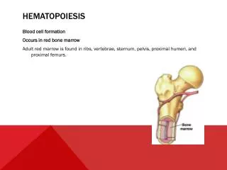
Hematology 425 Hematopoiesis. Russ Morrison September 18, 2006. Peripheral Smear Examination – Preparation of Smear. Review Chapter 1 of Atlas Properly prepared blood smear is essential to accurate assessment of cellular morphology Wedge smear preparation – well-prepared

E N D
Hematology 425 Hematopoiesis Russ Morrison September 18, 2006
Peripheral Smear Examination – Preparation of Smear • Review Chapter 1 of Atlas • Properly prepared blood smear is essential to accurate assessment of cellular morphology • Wedge smear preparation – well-prepared • 2/3 to ¾ of slide length covered by smear • Slightly rounded at featheredge • Lateral edges should be visible
Peripheral Smear Preparation • Smooth without irregularities, holes, streaks • Featheredge should have a “rainbow” appearance • The whole drop is picked up and spread • Figure 1-2 is the desired smear, examples of unacceptable smears are provided in figure 1-3
Peripheral Smear Staining • Purpose of staining is to identify cells and recognize morphology easily through the microscope • Most commonly used stains are Wright stain or Wright-Giemsa stain which both contain eosin and methylene blue • Stains which contain more than one dye are called polychrome stains as are the stains above
Peripheral Smear Staining • Cells are fixed to the glass slide via the methanol that is present in the stain • pH-dependent staining, optimal pH 6.4 • Buffered stain of appropriate pH • Free methylene blue is basic, stains acidic cellular components on the slide (RNA) blue • Free eosin is acidic and stains basic components (Hgb, Eo granules) red
Peripheral Smear Staining • Optimally stained smear characteristics: • RBCs pink to salmon in color • Nuclei dark blue to purple • Cytoplasmic granules of neutrophils are lilac • Cytoplasmic granules of eosinophils are red to orange • Cytoplasmic granules of basophils are dk blue to black • Area between cells should be clean (no ppt)
Peripheral Smear Staining • Best staining results are obtained from freshly made slides prepared within 2-3 hours of blood collection • Slides must be allowed to dry thoroughly before staining
Peripheral Smear Examination • Now that you have the perfect smear, stained well: • Begin scanning on low power (10x) to assess quality of the smear and stain • Look for abnormal red cell distribution (rouleaux, auto-agg), disproportionate number of large nucleated cells at the edges of the smear (make another smear for the later case) • Look for abnormal cells (blasts, reactive lymphocytes, parasites)
Peripheral Smear Examination • Using the 40x (high dry) objective, find an area of the smear where the RBCs are evenly distributed • Scan 8-10 fields in this area and determine the avg WBCs per field • Check the WBC count by multiplying this avg by 2000 (approx.WBC count/mm3) • Discrepancies between instrument count and estimate should be resolved (QC!)
Peripheral Smear Examination • Using the 100x (oil) objective, in same area of slide as above count 100 consecutive WBCs • Classify the WBCs • Report as a % • Use “battlement” track (Figure 1-6 of atlas) to minimize WBC distribution errors
Peripheral Smear Examination • WBC abnormalities (toxic changes, Dohle bodies, reactive lymphs, Auer rods, etc) also reported, if observed • If present, NRBCs are counted and reported as #/100WBCs • RBC,WBC and PLT morphology performed • Platelet estimate performed (avg PLTs in 10 fieldsX20,000), compare to machine count • All abnormal findings verified under the 100x objective
Morphology & Function of Cellular Components • Cells are the structural units that constitute living organisms • Most cells have three basic parts • Membrane • Cytoplasm • Nucleus • Each of these parts has components that assist in varied functions • Table 5-1 in the text is an excellent summary of cell components and functions
M & F, Cell Membrane • A semipermeable outer boundary separating the cellular components from the environment • Three basic functions • restricts/facilitates interchange of substances with the environment • Detects hormonal signals facilitating cell-to-cell recognition • Supports the blood groups, histocompatibility loci and receptors of cellular identity
M & F, Cell Membrane • Cellular membranes must be resilient and elastic • Achieves the above properties by being a fluid structure of globular proteins floating in lipids • Membrane proteins are mostly glycoproteins found floating in lipid bilayers
M & F, Cell Membrane Proteins • Two types of proteins in cell membranes • Integral proteins – may traverse the lipid bilayer and penetrate to the outside of the membrane. “Transmembrane” proteins provide communication and transportation between the cell’s interior and the external environment. • Peripheral proteins – found only on the cytoplasmic side, form the cell’s cytoskeleton. Attach to the cytoplasmic ends of integral proteins to form a reticular network that maintains structural integrity and holds integral proteins in place.
M & F, Cell Membrane Carbos • Occur in combination with proteins and lipids (glycoproteins and glycolipids) • The carbohydrate portion almost always extends beyond the outer cell surface • Gives the cell a carbo coat referred to as the glycocalyx • Carbos function in cell-to-cell recognition, provide a negative surface charge, surface receptor sites and cell adhesion capabilities. • Many of the RBC antigens are carbos, genes code for proteins (transferase enzymes).
M & F, Nucleus • The nucleus is made up of 3 components • Chromatin • Nuclear envelope • Nucleoli • the control center of the cell • The largest organelle in the cell • Made largely of DNA • Site of DNA replication and transcription • Responsible for cellular chemical reactions • Responsible for cellular reproduction • Stains blue because of its acidic nature
M & F, Nucleus • Chromatin • Consists of nucleic acids and proteins • Two types of protein • Histones, negatively charged • Nonhistones, positively charged • Two types of chromatin • Heterochromatin – genetically inactively, dense and darkly staining • Euchromatin – genetically active, diffuse,, loosely coiled and stains pale blue, RNA transcription is able to occur here
M & F, Nucleus • Nuclear Envelope 1. surrounds the nucleus 2. consists of an inner and outer membrane • Outer membrane is continuous with an extension of the endoplasmic reticulum, between the 2 membranes is a diaphragm 50 nm in thickness continuous with the lumen of endoplasmic reticulum. Nuclear pores penetrate the nuclear envelope allowing communication between the nucleus and cytoplasm. (#pores decrease as cell matures)
M & F, Nucleus • Nucleoli - the nucleus contains one to four nucleoli -these organelles contain a large amount of RNA and other proteins in a loose fibrillar form - the site for the synthesis of various forms of RNA, which is then transported through the nuclear pores for ribosomal assembly and protein synthesis
M & F, Cytoplasm • The cytoplasmic matrix is a homogenous continuous aqueous solution called cytosol • It is the environment in which the organelles join and function • The organelles of the cytoplasm will be discussed individually
M & F, Cytoplasm • Golgi Complex • A series of stacked, membrane-bound, flattened sacs referred to as cysternae • Involved in modifying, sorting and packaging macromolecules for secretion or delivery to other organelles • The “traffic cop” of the cell
M & F, Cytoplasm • Endoplasmic Reticulum • Lace-like network found throughout the cytoplasm • Specializes in making and transporting lipid and membrane proteins • Rough endoplasmic reticulum has ribosomes engaged in the synthesis of proteins on its outer surface • Smooth ER does not include ribosomes and may serve as a storage site for new proteins
M & F, Cytoplasm • Ribosomes • Small particles composed of near-equal amounts of protein and RNA • Found free in the cytoplasm, on the surface of RER, and in the nucleus and nucleoli of cells • May exist singly (monoribosome) or form chains (polyribosome) • The more ribosomes present within the cell, the darker blue the staining characteristics • Serve as the site of protein synthesis accomplished with the assistance of transfer RNA, for amino acid transport to the ribosome, and messenger RNA, which provides sequencing information for the amino acids.
M & F, Cytoplasm • Mitochondria • Responsible for the metabolic processes of energy-producing reactions and electron transfer – oxidative reactions. • The oxidative systems described within the mitochondria are the Krebs cycle, fatty acid cycle and the respiratory chain. • Also present in the mitochondria are proteins, phosphorylase, ribosomes and DNA. • Mitochondria are capable of self-replication and their number is directly related to the amount of energy required by the cell. • Do not accept color with routine stains
M & F, Cytoplasm • Lysosomes • Contain hydrolytic enzymes bound within a membrane and are involved in the cell’s intracellular digestive process • The membrane prevents the enzymes from attacking the protein, nucleic acids, mucopolysaccharides, lipids and glycogen within the cell itself • The hydrolitic enzymes become active when lysosomes bind to the phagocytic vacuole and the membrane ruptures, allowing the escape of the enzymes into the phagosome • May require special staining techniques
M & F, Cytoplasm • Microfilaments • Are solid structures approximately 5 nm in diameter consisting of actin and myosin proteins • These fibrils or groups of fibrils are located near the nuclear envelope or close to the nucleus and assist in cell division • Are present near the membrane to assist in cytoskeletal support and motility
M & F, Cytoplasm • Microtubules • Are approximately 25 nm in diameter and of variable length • Organized from tubulin through self-assembly • Have several functions 1. help maintain the cell’s shape and assists movement of some intracellular organelles 2. makes up the mitotic spindle fibers and the centrioles during mitosis • Not seen in blood smears
M & F, Cytoplasm • Centrioles • Paired structures consisting of nine bundles of three microtubules within each bundle • Serve as insertion points for the mitotic spindle fibers during metaphase and anaphase of mitosis
M & F, Review Questions • What component of the erythrocyte is most responsible for its cytoskeleton and cellular integrity? 1. Integral protein 2. Peripheral protein 3. Nucleus 4. Golgi complex
M & F, Review Questions • What cellular component of the erythrocyte is its control center? 1. Heterochromatin 2. Golgi complex 3. Nucleus 4. Cytoplasm
M & F, Review Questions • How does the genetically active portion of the nuclear chromatin look when stained with Wright's stain? 1. Condensed, darkly stained 2. Clumped, dark purple 3. Ropey, pink or red 4. Diffuse, uncondensed and pale blue
M & F, Review Questions • What cellular component "directs traffic" for smooth functioning? 1. Mitochondria 2. Ribosomes 3. Golgi complex 4. Nucleoli
M & F, Review Questions • If the cytoplasm of a cell is very basophilic or blue it contains: 1. No ribosomes 2. Large number of ribosomes 3. Small number of ribosomes 4. Only polyribosomes
M & F, Review Questions • Protein synthesis occurs in what part of a cell? 1. Ribosomes 2. Nucleus 3. Mitochondria 4. Lysosomes
M & F, Review Questions • What cellular component is responsible for energy production and metabolic processes? 1. Ribosomes 2. Nucleus 3. Mitochondria 4. Lysosomes
M & F, Review Questions • The granules of phagocytic white cells are composed of: 1. Microfilaments 2. Lysosomes 3. Microtubules 4. Centrioles
M & F, Review Questions • What is the purpose of the granules in phagocytic cells? 1. Maintain cell shape 2. Manufacture enzymes 3. Provide surface receptor sites 4. Digest foreign material such as microorganisms