Download
1 / 35
510 likes | 1.16k Vues
Physiology of the Digestive System. Functional Anatomy of the git. DR . AMEL EASSAWI DR. Sharique Ahmed Quadri. OBJECTIVES . To know the components of GIT and their functional significance. Emphasize the functional importance of four layers of GIT.

E N D
Physiology of the Digestive System Functional Anatomy of the git DR. AMEL EASSAWI DR. Sharique Ahmed Quadri
OBJECTIVES • To know the components of GIT and their functional significance. • Emphasize the functional importance of four layers of GIT. • Outline four basic digestive processes. • Recognize the importance of regulatory factors that controls digestive functions.
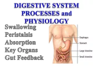
Digestive Tract Mouth Pharynx Esophagus Stomach Small intestine Duodenum Jejunum Ileum Large intestine Cecum Appendix Colon Rectum Anus Accessory Digestive Organs Salivary glands Exocrine pancreas Biliary system Liver Gallbladder Digestive System
Digestive System • Digestive tract is 4.5 m (15 feet) in normal contractile state. • Lumen is continuous from mouth to anus and hence is continuous with external environment. • Conditions that are essential for digestive process can be tolerated in the digestive tract lumen that could not be tolerated in body proper.
Digestive System Primary Function: • Transfer nutrients, water, and electrolytes from ingested food into body’s internal environment The Digestive System Performs Four Functions: • Motility • Secretion • Digestion • Absorption
Functions of the Digestive System Motility: • Muscular contractions that mix and move forward the contents of the digestive tract Two Types of Digestive Motility: • Propulsive (peristalsis) movements • Mixing (segmentation) movements
Functions of the Digestive System Propulsive Movements: • Push contents forward through the digestive tract • Velocity with which contents are moved forward (rate of propulsion) varies in different regions of GIT, depending on functions of that region. • Rapid movement in esophagus • Slow movement in small intestine
Functions of the Digestive System • Movements of contents through most of digestive tract is accomplished by contraction of smooth muscle except: • Mouth • Early part of esophagus • External anal sphincter • In these regions motility involves skeletal muscle (voluntary component)
Functions of the Digestive System Mixing Movements: • Serve Two Functions: • Mixing food with digestive juices & hence promotes digestion of foods. • Facilitates absorption by exposing all parts of intestinal contents to absorbing surfaces of digestive tract.
Functions of the Digestive System Secretions: • Digestive juices are secreted in to GIT lumen by exocrine glands. • Digestive secretion consist of: • Water • Electrolytes • Specific organic constituents(enzymes, bile salts, or mucus) important in digestive process.
Functions of the Digestive System • Secretions are released into the digestive tract lumen on appropriate neural or hormonal stimulation. • Normally reabsorbed in one form or another back into blood after their participation in digestion. Failure of reabsorption of digestive juices , as in diarrhea & vomiting results in loss of fluid
Functions of the Digestive System Digestion: • Biochemical breakdown of structurally complex foodstuffs into smaller, absorbable units by enzyme produce within GIT • Complex foodstuffs and their absorbable units • Carbohydrates → Monosaccharides (poly &disaccharides) (Glucose,fructose,galactose) • Proteins → Amino acids • Fats → Glycerol and Fatty acids (triglyceride)
Functions of the Digestive System Absorption: • In the small intestine, digestion is completed most absorption occurs. • Through process of digestion small absorbable units resulting from digestion, along with water, vitamins, and electrolytes are transferred from digestive tract lumen into blood or lymph.
Digestive Tract Wall • GIT wall has same general structure throughout length from esophagus to anus (with some local characteristic variations) • Four major tissue layers • Mucosa • Innermost layer • Submucosa • Muscularis externa • Serosa • Outer layer
Mucosa • Lines luminal surface of digestive tract • Highly folded surface greatly increases absorptive area • Three layers: • Mucous membrane • Lamina propria • Muscularis mucosa
Mucosa Mucous Membrane: • Inner epithelial layer serves as protective surface • Modified in particular areas for secretion and absorption. Contains: • Exocrine gland cells – secrete digestive juices • Endocrine gland cells – secrete blood-borne gastrointestinal hormones • Epithelial cells – specialized for absorbing digestive nutrients
Mucosa Lamina Propria • Middle layer of connective tissue on which epithelium rest. • Houses gut-associated lymphoid tissue (GALT) • Important in defense against disease-causing intestinal bacteria. MuscularisMucosa • Sparse layer of smooth muscle, contraction modifies the pattern of surface folding.
Submucosa • Thick layer of connective tissue. • Provides digestive tract with distensibility and elasticity. • Contains larger blood and lymph vessels • Contains nerve network known as submucosalplexus.
Muscularis Externa • Major smooth muscle coat of digestive tube • In most areas consists of two layers • Circular layer • Inner layer • Contraction decreases diameter of lumen • Longitudinal layer • Outer layer • Contraction shortens the tube • Together contractile activity of these layers produces propulsive and mixing movements • Myenteric plexus • Lies between the two muscle layers
Serosa • Outer connective tissue covering of GIT • Secretes serous fluid(watery, slippery fluid) • Lubricates and prevents friction between digestive organs and surrounding viscera. • Continuous with mesentery throughout much of the tract • This Attachment provides relative fixation • Supports digestive organs in proper place while still allowing them freedom for mixing and propulsive movements
Regulation of Digestive System Function • Digestive motility and secretion are carefully regulated to optimize the digestion. • Four factors are involved in regulating digestive system function. • Autonomous smooth muscle function • Intrinsic nerve plexuses • Extrinsic nerves • Gastrointestinal hormones
Autonomous smooth muscle function • In the wall of GIT some specialized smooth muscle cells are pacemakers cells –known as interstitial cells of Cajal. • These cells lie between circular & longitudinal layer of smooth muscles. • These are self excitable cell that displays rhythmic spontaneous variations in membrane potential-known as SLOW WAVE POTENTIAL OR basic electrical rhythm (BER).
Autonomous smooth muscle function • If slow wave reaches threshold, depolarization is triggered at the peak resulting in rhythmic cycles of contraction. • Whether threshold will reach or not depends on various mechanical, neural and hormonal factors that influence starting point of slow wave (e.g. presence of food bolus in GIT).
Autonomous smooth muscle function • The rate of self induced contractile activity depends on inherent rate established by involved pacemaker. • The intensity of contractions depends on number of action potentials occurring at peak of slow wave. • Greater the number of contraction--higher the cytosolic calcium--stronger the contraction.
Intrinsic nerve plexuses • Submucosal plexus and myentric plexus, together often termed as enteric nervous system. • Primarily coordinate local activity in GIT. • Intrinsic plexus can affect all functions of digestive tract, i.e. motility, secretion of digestive juices and gastrointestinal hormones. • Intrinsic nerve activity can be influence by endocrine, paracrine and nerve signals
Extrinsic Nerves • Are through both branches of ANS • Influence GIT motility secretion either by • Modifying activity of intrinsic plexuses. • Altering level of GI hormone secretion. • Directly acting on smooth muscle and glands. • Sympathetic inhibits the motility and secretion and parasympathetic increases both. • Extrinsic nervous system coordinate activity between different regions of GIT.
Gastrointestinal Hormones • Tucked within mucosa of certain regions of GIT are endocrine gland cells that releases hormone into blood on appropriate stimulation. • These hormones acts on other areas of GIT and exert either stimulatory or inhibitory influences on smooth muscle and exocrine cells.
References • Human Physiology, Lauralee Sherwood, seventh edition. • Text book Physiology by Guyton &Hall,11th edition. • Text book of Physiology by Linda S. Contanzo, third edition. • Physiology by Berne and Levy, sixth edition.