Ventilation
Ventilation. Intro: why do we breathe?. Key Terms. Ventilation : Movement of air into and out of the lungs Gas exchange : Movement of gases across membranes according to pressure gradients Pressure gradients : Determined by the partial pressure of the gas

Ventilation
E N D
Presentation Transcript
Key Terms • Ventilation: Movement of air into and out of the lungs • Gas exchange: Movement of gases across membranes according to pressure gradients • Pressure gradients: Determined by the partial pressure of the gas • Gases: Oxygen necessary for cellular respiration; Carbon dioxide is a volatile acid
Breathing, ventilation and respiration • Used synonymously • Used to think respiration occurred in the lung • Ventilation: movement of air • Respiration: cellular utilization of O2
Ventilation • Pulmonary minute ventilation (VE) • The rate of expired ventilation • Usually expressed in L/min • VE = VT x f • Expired ventilation and inspired essentially the same, may differ in transition from rest to exercise .
Environmental influences • Temperature, pressure, water vapor all impact gas volumes • Gas laws • Boyle’s law: Pressure and volume inversely related; P1V1=P2V2 • So, as pressure goes up, volume goes down and vice versa • Charles’ law: Temp and volume directly related; V1/T1 = V2/T2 • So, as temperature goes up, volume goes up and vice versa • Dalton’s law: The total P of a gas is determined by the partial pressures of all the constituent gases • STPD • Standard temperature, pressure, dry • ST=0°C, P=760 mmHg, 0 mmHg H2O vapor pressure (PH2O) • BTPS • Body temperature, pressure, saturated • Body temperature, P= ambient pressure, PH20=47 mmHg at 37°C • ATPS • Ambient temperature, pressure, saturated • T=ambient, P=ambient, PH20=47 mmHg at 37°C • Typically you collect at ATPS and convert to BTPS (ventilation) or STPD (Vo2, Vco2); allows comparison across studies
Entry of O2 into the blood • Determined Entirely by pressure gradients • Partial pressure • Pressure exerted by each gas in a composition • Atmospheric pressure (PAtm): 760 mmHg • Partial pressure of O2 (Po2): 159 mmHg (.2094 x 760) • Rest is nitrogen, some argon and very little CO2 • When air reaches alveoli, Po2 falls, why? • Think of gas laws
Entry of O2 into the blood 1) Water vapor • Gas is fully humidified, so at normal body temp, water vapor pressure is 47 mmHg 2) Co2 is also higher in the alveoli • Thus, Po2 of alveoli about 100-105 mmHg • 760 – 47 = 713 or the pressure of the air in the lung • Dalton’s Law • 713 x .2094 = 149 (inspired pressure of O2; PiO2) • PAO2 = PiO2 – (PACO2/RER) • Alveolar gas equation • PAO2 = 149 – (40/.85) or 149-47 = 102 • RER = respiratory exchange ratio or Vco2/Vo2 • Usually about 0.85 at rest with mixed diet
Entry of O2 into the blood • Once O2 gets into alveoli it diffuses into the blood • Due to favorable oxygen gradient (~100 to 40 mmHg) • Most binds with Hb (~97%) • Some dissolved in plasma (3%) • Oxygen content of blood (CaO2) CaO2=1.34*[Hb]*(%sat of Hb) + 0.003 * PaO2 CaO2 = 1.34 * (15mg/dl)*(.98) + 0.003* (100mmHg) CaO2 = ~20 ml/dl [Hb]= hemoglobin concentration PaO2 = partial pressure of oxygen
Pulmonary diffusion • Diffusion of gases through tissues (gel) • Major determinants • Partial pressure difference (major) • Solubility of the gas (minor) • Gases of lower solubility typically have greater partial pressure gradients
Rate of diffusion • Determined by • Area available • Thickness • Partial pressure gradient (P1-P2) • Diffusion coefficient • Determined by solubility and molecular weight
Rate of diffusion • CO2 is slightly larger than O2 (MW; 44 vs 32 g/mol) • CO2 has a much higher solubility coefficient (0.57 vs 0.024) • Thus, CO2 has a greater relative diffusion coefficient (~20 x higher) • Thus, O2 needs a larger pressure gradient to “force” itself across biological membranes
Arterial blood gas homeostasis • Maintenance of blood gases (PaO2 and PaCO2) very important • Keep driving pressure for CO2 and O2 high • Driving pressure is the difference between arterial and venous pressure (PaO2-PvO2) • Note that gradients increase with exercise
Oxygen transport • Oxygen content • CaO2 = 1.34[Hb]*(%sat) + 0.003 * PaO2 • Cardiac output (Qc) = HR * stroke volume • Thus, total oxygen transport capacity (or delivery) is Qc*CaO2 or Qo2 • Qo2 is a measure of how much oxygen is circulated around by the heart in one minute • So, if CaO2 = 20 ml/dl and Qc equals 30 L/min • Qo2 = 30 * 0.2 or • Qo2 = 6L/min
Shifting of O2 dissociation curve • Remember: we noted that exercise increases the pressure gradients • How?: O2 dissociation curve shifts • Curve shows the relationship between Po2, CaO2 and % Hb saturation • Right shifting increases O2 unloading • Right shift called Bohr effect • What shifts the curve?
Effects of Co2 and pH on O2 transport • The shape of the O2 dissociation curve is altered by 4 variables • pH • < 7.4 = right shift • >7.4 = left shift • Temperature • >38C = right shift • <38C = left shift • Co2 • >40 mmHg = right shift • <40 mmHg = left shift • 2,3 DPG (diphosphoglycerate) • Altitude increases this
Co2 transport • Co2 must be transported from tissues to blood and lungs for removal • Carried in 3 ways • Bound to Hb (carbamino compounds) (15-20%) • Dissolved in plasma (5-10%) • As bicarbonate (HCO3-), ~70%
Co2 transport • More Co2 dissolves (than O2) in plasma due to greater solubility • Binding of Co2 to Hb occurs at different site than O2 • Co2 combines with H2O to form bicarbonate
Co2 content • Amount of Co2 carried in the blood depends upon Pco2 • Unlike oxygen, the Co2 curve is linear over a much greater range • Thus, as Co2 production increases • greater driving pressure (from tissue to blood) • As Co2 is extremely soluble, this increases Co2 transport (No upper limit)
Effect of O2 on Co2 transport (and vice versa) • When Co2 increases in blood • Shifts O2 curve to right • Facilitates unloading of O2 at the tissues • Called Bohr effect • When O2 falls • Shifts Co2 curve up and right • Facilitates greater Co2 loading • Called Haldane effect • Thus, at the level of the tissue, high CO2 facilitates unloading of O2 which allows greater amount of CO2 to be carried in blood • At the lung, high O2 forces CO2 from Hb (and plasma) and it is then exhaled
Arterial blood gases • Note how ventilation and PaCo2 inversely mirror each other • Note also the effect on pH • Major function of the ventilatory system is to rid the body of Co2 and control pH • VA = VCo2/PaCo2
Buffering of metabolic acids • pH is a measure if the acidity of the blood • Several sources of acid are during exercise • Lactic acid (HLa) • Carbon dioxide • These cause a fall in pH • Bicarbonate is a very effective buffer • A buffer helps to prevent a change in pH pK: Dissociation constant. pH at which acid (or base) is 50% dissociated (50% acid and 50% base)
Buffering of metabolic acids • Lactic acid produced • HLa → La- + H+ • H+ + HCO3- → H2CO3 → H2O + CO2 (exhaled) • Co2 produced • CO2 + H2O → H2CO3 → HCO3- + H+ (reverses at lung) • pH • Negative logarithm of the hydrogen concentration • pH = pk for HCO3-+ log [base/acid) • pH = 6.1 + log [HCO3-/(pCO2 *0.03)] (Henderson-Hasselbalch eq.) • pH = 6.1 + log [24 /1.2) • pH = 6.1 + 1.3 • pH = 7.4
Control of pH • Co2 and pH (actually the H+) stimulate ventilation • Chemoreceptors • Carotid sinus • Centrally (medulla) • Sensitive to changes in Pco2 and H+ • Stimulate breathing to expel CO2 and partially compensate for the metabolic acidosis
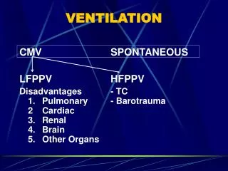
Ventilation • Gross Anatomy • Pharynx • Trachea • Bronchus • Alveolus
Ventilation • Ventilation • Moves air into and out of lung • Two separate areas of lung • Conducting zone • Respiratory zone • Conducting zone • Network of tubes whose function is movement of air • Trachea and Bronchi • Respiratory zone • Large, thin area where gas exchange occurs • Respiratory bronchioles and alveolar ducts
Ventilatory mechanics • Diaphragm • Main muscle of ventilation • Only skeletal muscle necessary for life • Accessory muscles • Intercostals • External • Inspiration • Internal • Expiration • Sternocleidomastoid, Scalenes • Inspiration • Abdominal muscles • Expiration
Ventilatory volumes • Note how much ventilation can increase • Due to large increases in tidal volume and frequency • Increases in tidal volume (VT) largely due to accessory muscles • Increases in frequency (f) due to diaphragm
Dead space and alveolar ventilation • Ventilation (VE) is the total amount of air moved in and out of the lungs • VE = VDS + VA • Dead space (VDS) • Anatomic dead space • Conducting zone • Physiologic dead space • Diseased areas • Dead space/tidal volume ratio • At rest ratio of VD/VT ~25-40% • With exercise VD/VT falls, why? • Alveolar ventilation • Ventilation of the gas exchange units
Static lung volumes • Volumes and capacities • Volume: single measure • Residual volume (RV) • The amount of air in the lung after a maximal expiration • Expiratory reserve volume (ERV) • The amount by which you can increase expiration after a normal exhalation • Inspiratory reserve volume (IRV) • The amount by which you can increase inspiration after a normal inspiration • Tidal volume (VT) • The volume of a normal breath • Total lung capacity (TLC) • RV, ERV, VT and IRV • Vital capacity • ERV, VT and IRV • Functional residual capacity • RV, ERV • Where humans breath from • Inspiratory capacity • VT, IRV
Composition of Alveolar gases Air breathing; no water or CO2 100% oxygen
Diffusion • Oxygen • Breathed into lungs • Diffuses across blood gas barrier • Binds with hemoglobin (97%) • Dissolved in plasma (3%) • Circulated to tissues • Diffuses into tissues • Binds with myoglobin • Keeps oxygen pressure homogeneous within tissues • Utilized in mitochondria Mb
Transit time • Capillary blood volume (Vc) • The blood that is in the capillaries at one instant in time • Transit time • the ratio of VC/blood flow • VC =~70 ml • Qc = 100 ml/s • TT = 0.7 sec • More than adequate for equilibration of blood gases • Note that CO2 equillibrates MUCH faster than O2; why?
Control of ventilation • Respiratory control center • Brainstem • Medulla • Pons • Feed forward • Central command • Feedback • Peripheral and central chemoreceptors
Central and peripheral control • Feed forward • Sometimes called “central command” • Co-activation of cardiovascular, ventilatory and musculoskeletal systems • Central chemoreceptors • Sensitive to changes in pH • Caused by Co2 as H+ cannot cross Blood brain barrier • CO2 + H2O H2CO3 HCO3- + H+ • Peripheral chemoreceptors • Carotid sinus • Muscle metaboreceptors • Both sensitive to changes in pH, PCO2 and PO2 (particularly at high atltitude) • Peripheral mechanoreceptors • Sensitive to limb movement
Feed forward 1 1 Peripheral3 Peripheral3 Central 2 3 & 4 Peripheral chemoreceptors and mechanoreceptors