Download
1 / 23
230 likes | 429 Vues
Irradiation facilities for semiconductor detectors and electronics at the INFN National Laboratory of Legnaro Andrea Candelori Istituto Nazionale di Fisica Nucleare and Dipartimento di Fisica, Padova. OUTLINE. • The SIRAD irradiation facility at the TANDEM accelerator:

E N D
Irradiation facilities for semiconductor detectors and electronics at the INFN National Laboratory of Legnaro Andrea Candelori Istituto Nazionale di Fisica Nucleare and Dipartimento di Fisica, Padova
OUTLINE • The SIRAD irradiation facility at the TANDEM accelerator: - high energy protons and ions. • The CN accelerator: - protons and neutrons. • Total dose tests: - Tungsten (W) and Molybdenum (Mo) X-rays; - 60Co -rays.
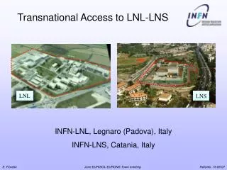
The SIRAD Irradiation Facility The SIRAD irradiation facility is located at the Tandem accelerator of the INFN National Laboratory of Legnaro (Padova, Italy). Tandem accelerator: -Van de Graaff type; 15 MV maximum voltage; two strippers; -servicing 3 experimental halls for nuclear and interdisciplinary Physics; Schematics of the 15 MV Tandem Van de Graaff accelerator and of the SIRAD irradiation facility at the +70º beam line (left). A photograph of the SIRAD irradiation facility is also shown for completeness (right).
Typical ion species available at SIRAD •Ionspecies from1H(22-30 MeV) up to197Au(1.4 MeV/a.m.u.) •LET from0.02 MeVcm2/mg(1H) up to81.7 MeVcm2/mg(197Au) The energy values refer to the most probable q1 and q2 charge state, with two stripper stations, and the Tandem operating at 14 MV. 1st multi-source 2nd multi-source
Low flux (102-105 ions/cm2s) irradiation set-up on 22 cm2 The on-line beam monitoring system for defocused beams by the fixed and mobile diodes: -left: side view of the experimental set-up; -right: front view (transverse to the beam) of the fixed and mobile diode boards. The mobile diodes are mounted on the sample holder with the DUT. The figure is not drawn to scale.
High flux (>108-109 ions/cm2s) irradiation set-up on 55 cm2 The on-line beam monitoring for rastered proton and ion beams by the 33 battery of Faraday cups positioned behind the DUT: side view of the experimental setup. The aperture of each Faraday cup is 0.60.6 cm2. The figure is not drawn to scale.
Example of validation for space mission Validation of the ASIC for the GLAST Large Area Telescope • GLAST space telescope • International collaboration (NASA, ESA, ASI, INFN,...) • INFN Padova: radiation tests of tracker, DAQ electronics • ASICs validated for SEE at SIRAD • ASICs validated for TD at CNR-ISOF 60Co -ray source • COTS validated for SEE at SIRAD
Summary of the main research activities at SIRAD SEE in FPGA Charge loss in Flash E2PROM SEE in ASICs for CMS, GLAST, AGILE, ALICE SEB, SEGR in power MOSFETs RILC and RSB (Ultra-thin gate oxide) Silicon detectors More than 61 papers published in the last 4 years.
BERGAMO VIAREGGIO CASSINO PADOVA PAVIA SIRAD Collaboration in Italy and abroad 1) Dip. di Fisica and INFN Padova 2) INFN Laboratori Nazionali di Legnaro 3) Dip. Ingegneria dell’Informazione, Padova 4) Tecnomare SpA (Venezia) 5) Center for Advance Space Optics (Trieste) 6) Dip. Fisica and INFN, Trieste 7) ITC-IRST (Trento) 8) Dip. Informatica e Telecomunicazioni, Trento 9) INAF, Sezione di Milano 10)ST Microelectronics (Agrate Brianza, Milano) 11) Dip. Elettronica, Pavia 12) Dip. Ingegneria Industriale, Bergamo 13) Dipartimento di Fisica Sperimentale, Torino 14) Dip. Automatica e Informatica,Politecnico di Torino 15) Dip Fisica and INFN, Bologna 16) Dip. Energetica and INFN, Firenze 17) Aurelia Microelettronica S.p.A. (Viareggio) 18) Dip.Ingegneria Elettronica, Università Roma 2 19) INAF, Sezione di Roma 20) DAEIMI e DSM, Università di Cassino 21) ST Microelectronics (Catania) A) Institut für Experimentalphysik (Amburgo, Germania) B) LETI (Grenoble, Francia) C) Centro Nacional de Microelectronica (Barcellona, Spagna) D) IMEC (Lovanio, Belgio) E) Philips Semiconductor (Nijmegen, Olanda) F) CERN (Ginevra, Svizzera) G) Helsinki Institute of Physics (Finland) H) Santa Cruz Institute for Particle Physica (California, U.S.A)
CN accelerator Characteristics: Van de Graaff type, 7 MV maximum voltage; Ion species: p (1H); d (2H); t (3H); 4He (single or double charge)and 15N (double charge) Max energy: 7 MeV for single charged species;14 MeV for 4He++; 8 MeV for 15N++. T(d,n)4He 9Be(d,n)10B with moderator D(d,n)3He 7Li(p,n)7Be 9Be(d,n)10B
X-ray tube Y axis motor Laser pointer Semi-automatic probestation Z Y X W and Mo X-rays: Seifert Rp-149 Irradiation Facility • Tube with W (7.4-12.06 keV L-lines) or Mo (17.4-19.6 keV K-lines) anode. • Maximum tube voltage 60 kV. Maximum tube current 50 mA. • X,Y (motorized) and Z (manual) axis for accurate position setting of the tube. • Radiation hardness qualification of the APV25 chip for the CMS silicon tracker.
140 D=10 cm D=15 cm 120 D=20 cm D=40 cm 13.9 mm 100 80 Dose rate (rad(Si)/s) 60 15.5 mm 40 16.1 mm 20 20.6 mm 0 -20 -15 -10 -5 0 5 10 15 20 140 D=10 cm D=15 cm 120 D=20 cm D=40 cm 7.2 mm 100 80 Dose rate (rad(Si)/s) 60 9.2 mm 40 11.4 mm 20 19.4 mm 0 -20 -15 -10 -5 0 5 10 15 20 W and Mo X-rays: radiation field dimensions X position (mm) Y position (mm)
60Co -ray source (CNR-ISOF) • Irradiation Facility: Panoramic Gammabeam model 150 A produced by Nordion Ltd (Canada) • Photon energies: 1.165 MeV and 1.332 MeV • Present activity: 2000 Ci ( 7.41013 Bq) • Point source for D>10 cm (D=10-300 cm) • Dose rate: ~5 rad(Si)/s at D=20 cm, ~1 rad(Si)/s at D=45 cm
Conclusions • The SIRAD irradiation facility at the 15 MV TANDEM accelerator: - Ion species from1H (23-30 MeV) up to197Au (1.4 MeV/a.m.u.) - LET from 0.02 MeVcm2/mg up to 81.7 MeVcm2/mg - High (>108-109 ions/cm2s) and low (102-106ions/cm2s) flux set-up - Ion Electron Emission Microscopy possibility - New irradiation chamber and sample holder (ESA standards) • The CN accelerator: - Monochromaticspectra: D(d,n)3He, T(d,n)4He, 7Li(p,n)7Be - Continuousspectra:9Be(d,n)10B - Thermal neutrons:9Be(d,n)10B with moderator • Total dose tests: - X-rays: W (L-lines at 7-12 keV) and Mo (K-lines at 17-20 keV) anode; dose rate: 120 rad(Si)/s. - -rays: 60Co with 1-5 rad(Si)/s dose rate (D=20-45 cm).
Scuola Nazionale “Rivelatori ed elettronica per applicazioni spaziali, Astrofisica e Fisica delle Alte Energie” INFN Laboratori Nazionali di Legnaro 4-8 Aprile 2005 More information on the web site http://sirad.pd.infn.it/scuola_legnaro
What is SIRAD? SIRADis the acronym forSIlicon andRADiation. The SIRAD irradiation facility is dedicated: "to investigate radiation effects on silicon detectors, electronic devices and systems in radiation hostile environments". -Total dose effects as a result of ionization damage. -Bulk effects as a result of displacement damage. -Single event effects as a result of an energetic particle strike. -High energy physics experiments. -Space missions of scientific and commercial satellites.
SIRAD upgrade: the Ion Electron Emission Microscope (IEEM) Purpose: Single Event Effect mapping, Ion Beam Induced Charge Collection studies. Nuclear Microprobe: µ-focused beam IEEM: defocused beam (Sandia) Ion beam 2D electron detector at focal plane of electron optics Object slit (Xhit,Yhit) Nuclear Microprobe: magnet optics for focusing (e.g. triplet) and electron optics for scanning secondary electrons Ion beam electron optics (Xbeam,Ybeam) coating rastering pattern channeltron hit confirmation by secondary electrons analysis of signal analysis of signal target target Resolution on target determined by beam optics spot size and positioning. Resolution on target: lateral size of field of view divided by linear line pair resolution of sensor.
SIRAD upgrade: the Ion Electron Emission Microscope (IEEM) UV lamp (PEEM) SIRAD contrast diaphragm I I lens PSD Image intensifier The ion impact position on the target is determined by “imaging” the position from which secondary electrons are emitted: the intrinsic resolution is of the order of 0.6 m over a 250 m field of view.
IEEM images with UV lamp and ion beam • Lattice step: 40 mm • Structure width is about 6 mm. • The lattice is made by copper. UV lamp 223 MeV Br ion beam
W anode, 50kV, 0.1 mm Al filtration 17.4 keV 7.6-12.06 keV Mo anode 30 kV, 0.1 mm Mo filtration Mo 19.6 keV W W and Mo X-rays: emission spectra 1.0 0.8 0.6 Photons/(mAsmm2) at 750 mm normalized to maximum 0.4 0.2 0.0 0 5 10 15 20 25 30 35 40 45 50 Photon energy (keV)