Heart Failure 101
Heart Failure 101. Armaan Khalid. What is Heart Failure?. HF is a syndrome that manifests as the inability of the heart to fill with or eject blood HF can result from any structural/functional cardiac disorder that impairs the ability of the heart to function normally

Heart Failure 101
E N D
Presentation Transcript
Heart Failure 101 Armaan Khalid
What is Heart Failure? • HF is a syndrome that manifests as the inability of the heart to fill with or eject blood • HF can result from any structural/functional cardiac disorder that impairs the ability of the heart to function normally • Coronary Artery Disease (CAD) is the most commonest cause of HF • Anything that ↑ myocardial work may aggravate/initiate HF
Common Causes of HF • Ischaemic Heart Disease (35-40%) • Cardiomyopathy (dilated) (30-34%) • Hypertension (15-20%)
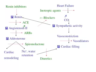
Pathophysiology • EXTREMELY COMPLEX! • Compensatory physiological changes that eventually get overwhelmed & become pathological • Factors involved are: • Venous return (preload) • Outflow resistance (afterload) • Myocardial contractility (inotropic state) • Salt & water retention
Frank-Starling’s Law of the Heart • Stroke work is increased as ventricular end diastolic volume is raised (e.g. ↓ ejection fraction) • ↑ Preload will ↑ cardiac contractility • Compensatory mechanism
Stroke Volume • SV is the volume of blood pumped from one ventricle of the heart with each heart beat • Usually assumed to be the Left • SV = EDV – ESV • Ejection Fraction = SV / EDV • Determinants • Preload • SV is controlled by preload due to Frank-Starling’s Law • Afterload • ↑ Afterload will ↓ SV • Contractility • ↑ Ca2+
Venous Return (Preload) • In HF, ejection fraction ↓ • Can be compensated by ↑ heart rate (sinus tachycardia) • In severe myocardial dysfunction, cardiac output can only be maintained by ↑ venous pressure (Preload) &/ ↑ tachycardia • Low functional reserve • Perfusion only maintained to vital organs (huge impact) • Causes dyspnoea, hepatomegaly, ascites, oedema • Due to ↑ venous pressure
Outflow Resistance (Afterload) • Afterload is defined as the myocardial wall tension developed during systolic ejection • It is formed by: • Pulmonary & systemic resistance • Physical characteristics of the vessel walls • Volume of blood that is ejected • ↑ in afterload ↓ cardiac output • ↑ end-diastolic volume • ↑ dilatation of the ventricle • ↑ AFTERLOAD • Vicious cycle
Myocardial Contractility • Sympathetic nervous system is activated in early HF as a compensatory mechanism • Inotropic support & maintains cardiac output • Chronic sympathetic activation leads to ↑ neurohormonal activation & myocyte apoptosis • Also causes ↑ cytosolic Ca2+ entry • Augments myocardial contractility • Impairs myocardial relaxation (lusitropy)
Salt & Water Retention • ↓ CO leads to diminished renal perfusion • Activation of RAAS • ↑ Aldosterone production to retain salt & water • Exacerbates increased venous pressure
Myocardial Remodelling • Primary response to chronic ↑ wall stress is myocyte hypertrophy, apoptosis & regeneration • Myocardial remodelling is pathological (eccentric) • Worsens the situation • ↑ stress on remaining myocytes
Findings Left Heart Failure Right Heart Failure • Clinical features • Fatigue, dyspnoea • Cardiomegaly • On auscultation, gallop rhythm • Crackles in lung bases • Pulmonary oedema • Clinical features • Fatigue, dyspnoea, anorexia, nausea • Jugular venous distension • Hepatomegaly • Pitting oedema • Ascites • Pleural transudates
Workup • FBE/LFT/U&E/TFT/Cardiac troponins • CXR (to be discussed) • ECG • Signs of ischaemia, MI, ventricular hypertrophy, LBBB • Echo (TTE/TOE) • ? Stress • BNP • Highly indicative of CHF & poor prognosis factor • ? Cardiac Biopsy • ? Cath Lab
General Lifestyle Advice • Educate • Obesity control • Dietary modification • Low salt, minimise EToH +/- fluid restriction • Smoking • Sexual activity • Exercise • Light exercise is encouraged • Vaccination
Radiological Findings in CHF • Presents typically as 1 of 2 radiographic patterns: • Pulmonary interstitial oedema • Pulmonary alveolar oedema • Which radiographic pattern appears depends on the pulmonary (venous) capillary wedge pressure (PCWP)
Pulmonary Interstitial Oedema • 4 key radiographic signs: • Thickening of the interlobular septa • Kerley B lines • Named after Irish neurologist & radiologist, Peter James Kerley • Peribronchial cuffing • Fluid in the fissures • Pleural effusions
Kerley B Lines • Thickening of the interlobular septa • Not visible on normal CXR • Only visible when it accumulates excessive fluid, PCWP about 15 mm Hg • Visible on frontal CXR, @ lung bases, @/ near costophrenic angles • Very short (1-2cm), very thin (1mm) & horizontal in orientation • Chronic Kerley B Lines • After repeated episodes of pulmonary interstitial oedema, fibrosis occurs
Kerley A Lines • Yes, there are A & C lines unfortunately • Kerley A Lines • Appears when connective tissue around the bronchoarterial sheaths in the lungs distends with fluid • Extends from the hila (up to 6 cm) & don’t extend to the lung peripheries • Kerley C Lines • If you know what they are, you are wasting time in this lecture • ? Overlap b/w A & B Lines • ? Myth
Peribronchial Cuffing • Bronchi may only be visible when seen on-end in the region of the pulmonary hila • Anywhere else, it is pathological • Fluid collects in the interstitial tissue surrounding the wall of the bronchi • Bronchial wall becomes ‘thicker’ & appears as doughnuts when seen on-end • Same mechanics as air bronchograms
Fluids in the Fissures • The fissures may normally be visible however, are almost never thicker than a line drawn with a sharp pencil • Fluid may collect b/w the 2 layers of the visceral pleura or subpleural space • Accumulated fluid distends the fissure(s) • Thicker, irregular & more visible
Pleural Effusions • Pleural effusions caused by CHF are usually bilateral • When it is unilateral, almost always right-sided • Therefore when you see a left-sided pleural effusion, consider • Mets, TB, thromboembolic disease, etc • Laminar Pleural Effusion • Thin, band-like density along lat chest wall, esp. near costophrenic angles (still sharp) • Almost always due to left atrial pressure ↑↑↑ • CHF • Lymphangitic spread of malignancy
Pulmonary Alveolar Oedema • Fluid spills out of the interstitium & into the airspaces when PCWP is sufficiently ↑↑↑ (25 mm Hg) • Almost known as Pulmonary Oedema • Radiographic finding • Fluffy, indistinct patchy airspace densities • Outer 1/3 usually spared • Lower zones > Upper zones • Butterfly/Bat-wing appearance • Pleural effusions usually present when the oedema is cardiogenic in nature
Emergency!!! • Mr XY, 80 y/o pensioner • HOPC sudden & extreme SOB • Wheezing & diaphoretic • Coughing with pink frothy sputum • Cold peripheries • Gallop rhythm • Differentials • Investigations • Management
Differentials??? • Asthma • COPD • Pneumonia • Pulmonary oedema • All of the above
Investigation!!! • CXR • ECG • U&E, Cardiac Troponins, ABG • BNP • Echo
Management • Sit upright & Oxygen • IV access & ECG monitoring • Morphine (5-10mg) +/- metoclopramide (10mg) • Beware of morphine systolic BP < 90 mm Hg • IV diuretics (furosemide 40-80 mg slowly) • GTN (spray 2 puffs/ 2 x 0.3 mg SL) • Don’t give if systolic BP < 90 mm Hg • Consider nitrate infusion if systolic BP > 100 mm Hg • Isosorbide dinitrate 2-10mg/h IV • Evaluate situation • If worsening, consider more diuretics & venesection • Get HELP!!!