Download
1 / 77
820 likes | 1.53k Vues
Arterial Blood Gases. Interpretation of arterial blood gases. Check Machine Clinical History Oxygenation Ventilation Acid base status. Is the Machine correct ?. H x HCO3 = 24 PCO2 pH 7.4 H+ = 80 – 40 = 40 pH 7.3 H+ = 80 – 30 = 50 pH 7.5 H+ = 80 – 50 = 30. Clinical History.

E N D
Interpretation of arterial blood gases • Check Machine • Clinical History • Oxygenation • Ventilation • Acid base status
Is the Machine correct ? • H x HCO3 = 24 PCO2 • pH 7.4 H+ = 80 – 40 = 40 • pH 7.3 H+ = 80 – 30 = 50 • pH 7.5 H+ = 80 – 50 = 30
Clinical History • Metabolic Acidosis – DM, Renal failure, muscle over activity, hypotension, diarrhea Diamox, Metformin , Alcoholism • Metabolic Alkalosis – Vomiting, RT aspiration, hypovolemia, diuretics, hypokalaemia, Bicarb administration • Respiratory Acidosis – COPD, muscular weakness, post-op • Respiratory Alkalosis – Tachypnoea, Sepsis, hepatic coma,
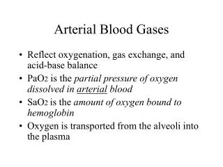
Oxygenation • Derived from PaO2 (partial pressure of oxygen in blood) and Saturation • PaO2- measured directly by the blood gas machine • Saturation- calculated value • Some ABG machines- in-built oximeter can give a directly measured value for saturation.
Oxygenation • A-a gradient – FiO2x(760-47)-PCO2x1.25 (normal 20) • Oxygen cost of breathing • Expected oxygen (PO2) = 500 x FiO2 • P/F ratio • SaO2 functional saturation OHbOHb+RHb
Ventilation & Acid-base status • Assessment of ventilation and acid base status go hand in hand • pH and PCO2- directly measured by the ABG machine • Bicarbonate and base excess- calculated values.
ABGnormal values • pH 7.4 (7.35-7.45) • PaCO2 35-45 mm Hg • HCO3- 22-26 mmol/l • BE +/- 5 • PaO2 >80(>60) mm Hg • pH & PaCO2 move in opposite directions • HCO3 & PaCO2 move in same direction
pH values and equivalent [H+] for water[H+] is the physical chemistry expression of molar concentration H+. • pH value [H+]; nmol.l)1-1 7.6 25 7.5 32 7.4 40 7.3 50 7.2 60 7.1 80 7.0 100 6.9 125 6.8 160
Base Excess • Actual Base Excess (ABE)– calculated by Van Slyke/Siggard Anderson equation (1948-50) • Standard Base Excess (SBE)– some base diffuses out of blood in vivo – correction does not take into account plasma protein & PCO2
Base Excess • The base excess is defined as the quantity of strong acid required to titrate blood to pH 7.40 with a PaCO2 of 40 mmHg (5.33 kPa) at 37 C • In practice, acid is not titrated as suggested but calculated using a variety of established formulae or normograms. • The base excess thus ‘removes’ the respiratory element of acid-base disturbance and identifies the metabolic contribution to interpret with pH and [H+]. • The standard bicarbonate is broadly similar and is the calculated [HCO3] at a PaCO2 of 40 mmHg [5.33 kPa]. • Although the base excess allows a metabolic acidosis to be diagnosed, it provides few clues as to the pathophysiology or underlying diagnosis.
Steps for Acid Base Status • Calculated Actual Anion Gap • Acidosis or Alkalosis • Respiratory or Metabolic • If Respiratory – Acute or Chronic • Metabolic Acidosis – Anion Gap • Other Metabolic Disorders • Respiratory Compensation for metabolic Disorders
When pH & PaCO2 change in same direction the primary problem is Metabolic • When pH & PaCO2 move in opposite directions & PaCO2 is normal the primary problem is Respiratory • If HCO3 & PaCO2 change in opposite direction then it is a mixed disorder (pH may be normal with abnormal PaCO2 or abnormal pH & normal PaCO2
Respiratory • The likely hood of complete compensation in chronic respiratory acidosis if PaCO2 > 60 is <15% • Acute compensation occurs within 6-24 hr • Chronic compensation occurs within 1-4 d • In clinical practice it is rare to see complete compensation. • Maxm compensation of pH 50-75% • In Ch respiratory alkalosis pH may be normal
COMPENSATIONS • Compensation in same direction dec. in pCO2 dec in HCO3 Acute Chronic • Inc. PaCO2 10 dec. pH 0.08 0.03 • Inc. PaCO2 10 inc HCO3 1 3 • dec. PaCO2 10 dec. HCO3 2 4 • Inc. HCO3 1 inc. PaCO2 0.5(0.5-1.0) • dec. HCO3 1 dec. PaCO2 1 lowest 7 -8
Ionic components of plasma including electrochemical equivalents • Principal cations (mEq.l)1-1) • Na (140) • K (4) • Ca (2) • Mg (2) • Total 148 mEq.l)1-1 • Principal anions (mEq.l)1-1) • Cl (100) • HCO3 (25) • Protein (15) • Phosphate specie (2) • Sulphate (1) • Organic acids (5) • Total 148 mEq.l)1-1
Anion Gap (1975) • Proteins – 15 K+ 4.5 • Org acids - 5 Ca+ 5.0 • Phosphates - 2 Mg+ 1.5 • Sulphates - 1 Total 23 11 Anion Gap = 23-11=12+4 Anion Gap = (Na+) - (HCO3 + Cl) Anion Gap with K+=8+4
Calculated Anion Gap • AG influenced by Albumin & pH • Change in Alb(4 gm) by 1 gm changes AG by 2 • Acidosis decreases AG by 2 • Alkalosis increases AG by 4 • Delta AG = AG – expected AG
Metabolic Acidosis • Metabolic acidosis is a non-respiratory process which has a tendency to produce a metabolic acidaemia, • The correct term when plasma pH < 7.35 ([H+] =45 nmol.l)1-1). • Strictly, during acidosis the pH may be in the normal range. • Clinically, a metabolic acidosis may be distinguished from a respiratory acidosis when alveolar hypoventilation is not the primary cause.
Metabolic Acidosis - Causes Normal Anion Gap • Diarrhoea • Intestinal/Pancreatic Fistula • Renal Tubular Acidosis • Fluids with high Chloride(DKA+NS) High Anion Gap • Lactic Acidosis • Ketoacidosis (diabetic, starvation) • Renal Failure • Poisoning (salicylates, ethanol, ethylene glycol)
Organic acids • Principally lactate and ketones. • Several thousand millimoles of lactate and ketones are metabolised per day (e.g. lactate 1500 mmol.day)1) traditionally attributed to the liver. • Kidney contributes a large component of the body’s metabolic disposal of lactate, perhaps up to 25–30%. • Hepatic urea production itself generates 2H+ for every molecule of urea produced
Inorganic acids • Sulphate and phosphate are the two most important & generated in the range of 1.5 mmol.kg)1.day)1-1. • Bioproducts of dietary protein and amino acid metabolism. • Sulphur-containing amino acids methionine and cysteine produce around 70% of the body’s total fixed acid per day in the form of sulphuric acid. • In chronic renal failure, sulphate may contribute up to 5 mEq.l)1 to the anion gap
Gap Gap Acidosis • Delta AG/Delta HCO3 • 1 = Met Acidosis with high AG • > 1.5 = Met Acidosis + Met Alkalosis • < 1 or 0 = Met Acidosis with normal AG
Metabolic Acidosis & Alkalosis • Gap-gap ratio > 1(AG:BE) (in presence of high AG acidosis when alkali is added the decrease in HCO3 is less than the increase in AG. In high AG Metabolic Acidosis gap-gap ratio >1 indicates co-existence of a Metabolic Alkalosis
Stewarts Physico-Chemical Approach • Sum of all +ve ions = sum of –ve ions • Aqueous solution is always neutral • Dissociated H+ exerts charge • Neutral pH – amount of dissociated H+ &OH- is equal at 25oC & 1 Atm pressure
Strong Ion Difference • Disossiates more in solution than weak ion • Strong cations – Na+, K+, Ca+, Mg+ • Weak cations – NH4, H+ • Strong anions – Cl-, Lactate • Weak anions – HCO3-, PO4-, OH- • As strong cation increases in solution the concentration of OH- increases more than the concentration of H+ ions – solution becomes more alkaline • Base Excess – change = metabolic • No change = respiratory
Stewart approach • SID – Blood has more strong cations (Na 135) than strong anions (Cl 100) – pH is more alkaline than water • Total Weak Acids –Albumin & Phosphates • Solutions with greater SID generate more HCO3 & vice versa • More alkaline the blood, the more HCO3 generated & the more acidic the blood the less HCO3 generated
Applications of Stewart’s Approach • Sepsis & Septic Shock – metabolic acidosis due to lactatemia • Hypoalbuminemia – metabolic alkalosis (can mask SID such as lactic acidemia) • Prolonged respiratory failure with associated hypercarbia leads to metabolic alkalosis because of Cl loss in urine
Applications of Stewart’s Approach • Mechanical ventilation increases the circulating volume of ANP & ADH resulting in increased total body water leading to dilutional acidosis • Renal failure causes metabolic acidosis. Polyuric renal failure may be associated with contraction alkolosis du to loss of Na, K, & free water.
Applications of Stewart’s Approach • Nasogastric suctioning causes hypochloremic alkalosis • Diarrhoea causes acidosis by loss of Na & K • Fever, sweating leads to insensible loss & contraction alkalosis • Antibiotics diluted in Na rich solutions increases SID & alkalosis
Applications of Stewart’s Approach • Lorazepam large volumes of propylene glycol cause metabolic acidosis • CRRT clears acidosis of renal failure by removing strong ions & phosphate unmasks metabolic acidosis due to hypovolemia • Loop diuretics cause hypochloremia & contraction alkalosis
Applications of Stewart’s Approach • Carbonic anhydrase inhibitors increase CO2 levels causing respiratory acidosis & cause diuresis leading to contraction alkalosis • Contraction alkalosis should be treated with free water • Hypochloremic alkalosis should be treated by correcting Cl deficit using NS
Applications of Stewart’s Approach • Mannitol causes dilutional acidosis. Contraction alkalosis follows due to diuresis • Normal & Hypertonic saline cause hyperchloremic acidosis • Diabetes insipidus can cause contraction alkalosis
Corrected HCO3 • >24 Alkalosis • <24 Acidosis • Actual HCO3 + delta AG
Urinary Anion Gap • In normal anion gap acidosis • (Na + K) – Cl (urinary pH < 6.5, no ketosis • Neg UAG = GI, iatrogenic • Positive UAG (> 20-30) – RTA type I, II, IV • Urinary pH > 6.0 = distal type I RTA • Urinary pH < 5.5 = proximal type II/IV RTA • Hypokalaemia = proximal type II RTA • Hyperkalaemia = Aldosterone deficiency type IV RTA
Metabolic Acidosis with decreased AG • Multiple Myeloma • Hypoalbunemia • Hyperkalaemia • Hypermagnesemia • Hypertriglyceredemia • Lithium Toxicity • Bromide Toxicity(pyridostigmine)
Metabolic Alkalosis • Urine Cl < 10 mEq/l – saline responsive • Urine Cl > 20 mEq/l – saline unresponsive
Urinary Anion Gap • Done in normal AG Metabolic Acidosis • (Na + K) – Cl • Neg – GI loss • > 20-30 – RTA type I, II, IV
Central Venous O2 Saturation (ScvO2) • ScvO2 most valuable in identifying trends in the balance of DO2 & VO2 • ScvO2 <70% identifies a state of inadequate O2 delivery relative to O2 consumption decreased DO2–low CO, Anemia, Hypoxia increased VO2 - hypermetabolism
2 Few Values • pH 7.30 PaCO2 60 PaO2 101 HCO3 28 • pH 7.228 PaCO2 45 PaO2 100 HCO3 18 • pH 7.55 PaCO2 26 PaO288 HCO3 28 • pH 7.50 PaCO2 40 PaO2 98 HCO3 36
A 60 year old male,known case of COPD was admitted with history of acute exacerbation of breathlessness. The ABG: pH- 7.204 PaCO2- 68 PaO2- 65 HCO3 28 • A 40 year old IDDM was admitted with breathless following Acute Gastritis.The ABG was : pH 7.28 PaCO2 18.4 PaO2 152 HCO3 8.5 Na 127 K 3.3 Cl 101 RBS 590mg% Urine Ketones -positive
A 40 year old female presented with acute breathlessness .Chest was clinically clear. She had similar attacks in the past. The ABG was pH 7.583 PaCO2 15.9 PaO2 137.9 HCO3 14.5 • A 25 years old boy was admitted along with severe vomiting following food poisoning. The ABG was : pH 7.52 PaCO2 45 PaO2 94 HCO3 32 Na 130 K 3.1 Cl 86
Expected changes in pH and HCO3- for a 10-mm Hg change in PaCO2 resulting from either primary hypoventilation (respiratory acidosis) or primary hyperventilation (respiratory alkalosis): ACUTE CHRONIC • Resp Acidosis pH ↓by 0.07 pH ↓ by 0.03 HCO3-↑ by 1* HCO3-↑ by 3 - 4 • Resp Alkalosis pH ↑ by 0.08 pH ↑ by 0.03 HCO3-↓ by 2 HCO3-↓ by 5 * Units for HCO3- are mEq/L
SKM a case of complicated Falciparum Malaria was admitted with SOB,decreased urination ,upper GI bleed & 2 episodes of sizzure.His ABG was pH 7.389 PaCO2 32.9 PaO2 153 5l/m of O2 HCO3 19.2 Urea 97.2 Creatinine 2.8 • He underwent HD pH 7.407 PaCO2 38.4 PaO2 52 5l/m of O2 HCO3 23. CXR ARDS . • He was on mech. Ventilation pH 7.406 PaCO2 34.7 PaO2 101 FiO2 o.5 HCO3 21.1 • 9 days later after his 6th HD,he had a fainting attack,detected to have tachycardia and hypotension pH 6.962 PaCO2 39.8 PaO2 271 FiO2 1.00 HCO3 8.8 • Immidiately ventilated and subsequently had malena • Post Bicarbonate pH 7.136 PaCO2 42.1 PaO2 273 FiO2 1.00 HCO3 10.8 • Post HD pH 7.462 PaCO2 37.8 PaO2 200 FiO2 1.00 HCO3 26.1 • Post extubation pH 7.442 PaCO2 52.9 PaO2 225 FiO2 0.80 HCO3 34.9 pH 7.570 PaCO2 24 PaO2 080 3l/m of 02 HCO3 21.3
Case 1 • A 28year female presented to the hospital with fever for 2days & Status Epilepticus. She had an cardiac arrest during a prolonged seizure & was immediately intubated, CPR was started, cardiac rhythm was restored & she was connected to a ventilator. Her ABG done was : • pH-6.788, pCO2-65,pO2-392(FiO2-1) • One hour later pH-7.175,pCO2-23,pO2-254(.8) • 7hours later pH-7.456,pCO2-24, pO2-300(.8)
Respiratory acidosis • PCO2 65, expected pH-7.2 • Actual pH 6.788 – metabolic acidosis • Cause – post-tictal lactic acidosis • Respiratory + Metabolic acidosis