Chapter 14 Endocrine System
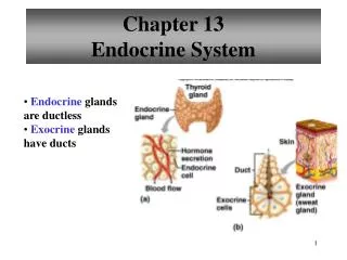
Chapter 14 Endocrine System. Introduction. The endocrine system and the nervous system are the two major communicating and coordinating systems in the body. The endocrine system communicates through chemical signals called hormones. Hormones. Classification of Hormones

Chapter 14 Endocrine System
E N D
Presentation Transcript
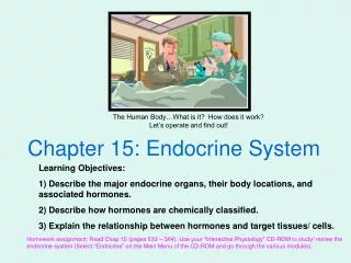
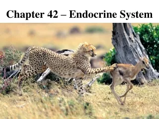
Introduction • The endocrine system and the nervous system are the two major communicating and coordinating systems in the body. The endocrine system communicates through chemical signals called hormones.
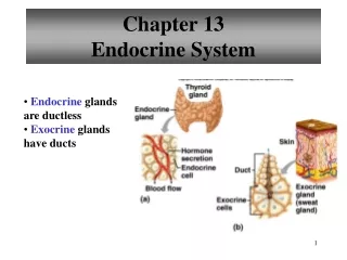
Hormones • Classification of Hormones • Hormones are secreted by endocrine glands directly into the blood. • Hormones are classified as proteins (protein-related substances) and steroids. • Hormone Receptors • Hormones are aimed at receptors of target organs. • Receptors are located on the outer surface of the membrane or inside the cell. • Hormone secretion is controlled by three mechanisms: negative feedback control, biorhythms, and control by the central nervous system.
Pituitary Gland • Hypothalamic-Hypophyseal Portal System • The portal system is a system of capillaries that connects the hypothalamus and the anterior pituitary. • The portal system transports releasing hormones from the hypothalamus to the anterior pituitary gland.
Pituitary Gland - cont’d • Hormones of the Anterior Pituitary Gland • Growth hormone stimulates growth and maintains blood glucose during periods of fasting. • Prolactin (lactogenic hormone) stimulates milk production by the breasts. • Tropic hormones stimulate other glands to secrete hormones. These include thyrotropin, adrenocorticotropic hormone, and the gonadotropins.
Pituitary Gland - cont’d • Hormones of the Anterior Pituitary Gland—cont’d • Thyroid-stimulating hormone stimulates the thyroid gland. • Adrenocorticotropic hormone (ACTH) stimulates the adrenal cortex. • The gonadotropic hormones stimulate the gonads (ovaries and testes).
Pituitary Gland - cont’d • Hormones of the Posterior Pituitary Gland • Antidiuretic hormone (ADH) stimulates the kidney to reabsorb water. • Oxytocin stimulates the uterine muscle to contract for labor and stimulates the breast to release milk during suckling (milk let-down reflex). • A tiny, third lobe secretes melanocyte-stimulating hormones.
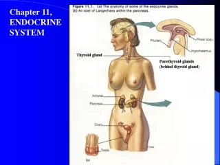
Other Endocrine Glands • Thyroid Gland • The follicular cells synthesize triiodothyronine (T3) and tetraiodothyronine, or thyroxine (T4). T3 and T4 regulate metabolic rate. • The parafollicular cells secrete calcitonin. Calcitonin lowers blood calcium. • Parathyroid Glands • The parathyroid glands secrete parathyroid hormone (PTH). • PTH stimulates the bones, kidneys, and intestines to increase blood calcium levels.
Other Endocrine Glands - cont’d • Adrenal Gland • The adrenal medulla secretes the catecholamines epinephrine and norepinephrine and causes the “fight or flight” response. • The adrenal cortex secretes the steroids: glucocorticoids, mineralocorticoid, and sex hormones. • Pancreas • The pancreas secretes insulin and glucagon. • Insulin lowers blood glucose while glucagon increases blood glucose.
Other Endocrine Glands - cont’d • Gonads • The ovaries are stimulated by the gonadotropins and secrete estrogens and progesterone. • The testes are stimulated by the gonadotropins and secrete testosterone. • Thymus Gland: plays an important role in the immune response • Pineal Gland: houses the “biological clock” and secretes melatonin • Other hormones include organ-specific hormones (cholecystokinin), prostaglandins, and hormones of adipose tissue
Introduction • The sensory system allows us to experience the world through a variety of sensations: touch, pressure, pain, proprioception, temperature, taste, smell, vision, hearing, and equilibrium.
Receptors and Sensation • Receptor • A receptor is a specialized area of a sensory neuron that detects a specific stimulus. • The five types of receptors are chemoreceptors, pain receptors (nociceptors), thermoreceptors, mechanoreceptors, and photoreceptors.
Receptors and Sensation - cont’d • Sensation • A sensation is a conscious awareness of incoming sensory information. • There are four components of a sensation. • The two characteristics of sensation are projection and adaptation.
General Senses • Pain • Pain receptors (nociceptors) are free nerve endings. • The stimuli for pain are tissue damage, lack of oxygen, and stretching or distortion of tissue.
General Senses - cont’d • Touch and Pressure • Receptors are mechanoreceptors and respond to forces that press, move, or deform tissue. • The receptors for pressure are located in the skin, subcutaneous tissue, and the deep tissue.
General Senses - cont’d • Temperature • There are thermoreceptors for heat and cold. • Thermoreceptors are found in free nerve endings and in other specialized sensory cells beneath the skin.
General Senses - cont’d • Proprioception • Proprioreceptors are located primarily in the muscles, tendons, and joints. • Proprioreceptors sense orientation or position.
Special Senses • Sense of Smell: The Nose • Olfactory receptors are chemoreceptors. • Sensory information travels along the olfactory nerve to the temporal lobe.
Special Senses - cont’d • Sense of Taste: The Tongue • Taste buds contain chemoreceptors for taste. • There are four basic taste sensations: sweet, salty, sour, and bitter. • Sensory information travels along the facial and glossopharyngeal nerves to the gustatory cortex in the parietal lobe.
Special Senses - cont’d • Sense of Sight: The Eye • The visual accessory organs include the eyebrows, eyelids, eyelashes, lacrimal apparatus, and extrinsic eye muscles. • The eyeball has three layers: the sclera, choroids, and retina (contains the photoreceptors, rods, and cones). • The eyeball has two cavities. One is a posterior cavity filled with vitreous humor, the other is an anterior cavity filled with aqueous humor. • There are two sets of eye muscles: extrinsic and intrinsic.
Special Senses - cont’d • Sense of Sight: The Eye—cont’d • The extrinsic eye muscles move the eyeball. • The intrinsic eye muscles control the size of the pupil and shape of the lens for refraction. • Light stimulates the photoreceptors. • The electrical signal is carried to the occipital lobe via the visual pathway. • Steps in seeing are summarized in Figure 13-1.
Special Senses - cont’d • Sense of Hearing: The Ear • There are three parts of the ear: the external ear, middle ear, and inner ear. • The middle ear contains the ossicles. • The inner ear structure concerned with hearing is the cochlea. It contains the hearing receptors and organ of Corti. • Hearing information is carried by the cochlear nerve to the temporal lobe. • Steps in hearing are summarized in Figure 13-16.
Special Senses - cont’d • Sense of Balance: The Ear • The receptors are mechanoreceptors located in the vestibule and the semicircular canals of the inner ear. • The receptors are activated when the head changes position. • Balance information travels along the vestibular nerve to many areas of the brain (cerebellum, midbrain, and temporal lobe).
Autonomic or Visceral Reflexes • What They Do: Autonomic reflexes regulate organ function • Pathway: The sequence is receptor activation, sensory input ( CNS), motor neuron response, and effector response
Organization and Function of the Autonomic Nervous System • Divisions of the ANS: There are two divisions. • Sympathetic nervous system, called “Fight or Flight.” • Parasympathetic Nervous System, called “Feed and Breed.” • Autonomic Terminology and Autonomic Pharmacology • Drugs that affect the sympathetic nervous system are called sympathomimetic and sympatholytic. • Drugs that affect the parasympathetic nervous system are called parasympathomimetic and parasympatholytic.
Organization and Function of the Autonomic Nervous System - cont’d • Autonomic Tone and Vasomotor Tone • Background firing of the ANS causes autonomic tone. • Background sympathetic stimulation of the blood vessels causes vasomotor tone.
ANS: Neurons • Numbers and Ganglia • Preganglionic fibers are fibers that extend from the CNS to the ganglia. • Postganglionic fibers are fibers that extend from the ganglia to the effector organ.
ANS: Neurons - cont’d • Neurons of the Sympathetic Nervous System • The SNS is called the thoracolumbar outflow. • The sympathetic ganglia are located in a chain close to the spinal cord; the chain is called paravertebral ganglia. • The adrenal medulla secretes hormones that mimic the SNS.
ANS: Neurons - cont’d • Neurons of the Parasympathetic Nervous System • The parasympathetic nervous system is called the craniosacral outflow. • Parasympathetic fibers travel with cranial nerves; most parasympathetics run with the vagus nerve CN X. • Naming Fibers and Neurotransmitters • Cholinergic fibers secrete acetylcholine (ACh). • Adrenergic fibers secrete norepinephrine (NE).
ANS: Neurons - cont’d • Neurotransmitters: Termination of Activity • ACh is degraded immediately by acetylcholinesterase. • NE activity is ended primarily by reuptake of the NE into the nerve terminal and by MAO activity within the nerve terminal.
Receptors of the Autonomic Nervous System • Cholinergic Receptors • These are activated by ACh. • There are two types: muscarinic and nicotinic (with subtypes). • Adrenergic Receptors • Activated by NE • There are two types: alpha and beta (with subtypes).
Receptors of the Autonomic Nervous System - cont’d • Receptor activation and blockade can be determined by examining Tables 12-1, 12-3, and 12-4. • Autonomic Receptors: “Doing Autonomic Pharmacology” • Clinical examples where drugs target autonomic receptors
Introduction • The brain, spinal cord, and peripheral nervous system act as a vast communication system. The spinal cord transmits information to and from the brain. The peripheral nervous system brings information to the CNS (its sensory role) and delivers information from the CNS to the periphery (its motor role).
What the Spinal Cord Is • The spinal cord is a tubelike structure located in the spinal cavity, extending from the foramen magnum (occipital bone) to L1 • Arrangement of Nervous Tissue • The gray matter is a centrally located, butterfly-shaped area. • The white matter is composed of myelinated fibers arranged in tracts. Ascending tracts are sensory tracts. Descending tracts are motor tracts.
What the Spinal Cord Is - cont’d • Arrangement of Nervous Tissue—cont’d • Spinal nerves are attached to the spinal cord. All spinal nerves are mixed (they contain sensory and motor fibers). • Sensory nerve fibers travel to the cord through the dorsal root. Motor nerve fibers travel in the ventral root.
What the Spinal Cord Does: Functions • The spinal cord relays both sensory and motor information. • The spinal cord acts as a major reflex center.
Reflexes • A reflex is an involuntary response to a stimulus. • The four components to a reflex are a sensory receptor; an afferent (sensory) neuron; an efferent (motor) neuron; and an effector organ.
Peripheral Nervous System • Nerve • A nerve is a group of neurons, blood vessels, and connective tissue. • There are sensory nerves, motor nerves, and mixed nerves.
Peripheral Nervous System - cont’d • Structural Classification of Nerves • A classification of nerves on the basis of structure divides nerves into cranial nerves and spinal nerves. There are 12 pairs of cranial nerves (Table 11-3) and 31 pairs of spinal nerves (Table 11-5). • Spinal nerves are sorted out at nerve plexuses. The three major plexuses are the cervical plexus, the brachial plexus, and the lumbosacral plexus. • A dermatome is the area of skin innervated by each spinal nerve.
Peripheral Nervous System - cont’d • Functional Classification of Nerves • Somatic afferent nerves carry sensory information to the CNS. • Somatic efferent nerves carry motor information to skeletal muscles. • Autonomic nerves carry motor information to the organs (viscera).
Introduction • The purpose of the nervous system is to bring information to the central nervous system, interpret the information, and enable the body to respond to the information.
The Nervous System: Overview • Divisions of the Nervous System • The central nervous system (CNS) includes the brain and the spinal cord. • The peripheral nervous system includes the nerves that connect the CNS with the rest of the body.
The Nervous System: Overview - cont’d • Cells That Make Up the Nervous System • Neuroglia (glia) support, protect, and nourish the neurons. • Neurons conduct the nerve impulse. • The three parts of a neuron are the dendrites, cell body, and axon.
The Nervous System: Overview - cont’d • Types of Neurons • Sensory, or afferent, neurons carry information toward the CNS. • Interneurons are located in the CNS (make connections). • Motor, or efferent, neurons carry information away from the CNS toward the periphery.
The Nervous System: Overview - cont’d • White Matter and Gray Matter • White matter is due to myelinated fibers. • Gray matter is composed primarily of cell bodies, interneurons, and unmyelinated fibers. • Clusters of cell bodies (gray matter) are called nuclei and ganglia.
The Neuron Carrying Information • Nerve Impulse • The electrical signal is called the action potential or nerve impulse. • The nerve impulse is due to the following changes in the neuron: polarization, depolarization, and repolarization. • The nerve impulse is due to flow of ions: polarization (outward flux of K+), depolarization (influx of Na+), and repolarization (outward flux of K+).
The Neuron Carrying Information - cont’d • Nerve Impulse—cont’d • The refractory period is the unresponsive period of the neuron. • The nerve impulse jumps from node to node as it travels along a myelinated fiber. Myelination increases the speed of the nerve impulse. • The nerve impulse causes the release of a neurotransmitter.
The Neuron Carrying Information - cont’d • Synapse • The synapse is a space between two neurons. • The nerve impulse of the first (presynaptic) neuron causes the release of neurotransmitter into the synaptic cleft. The neurotransmitter diffuses across the synaptic cleft and binds to the receptors on the second (postsynaptic) membrane. The activation of the receptors stimulates a nerve impulse in the second neuron.
Brain: Structure and Function • Cerebrum • The right and left hemispheres are joined by the corpus callosum. • The four main cerebral lobes are the frontal, parietal, temporal, and occipital lobes. Functions of each lobe are summarized in Table 10-2. • Large areas of the cerebrum, called association areas, are concerned with interpreting, integrating, and analyzing information.