Download
1 / 60
640 likes | 810 Vues
Glomerular Diseases : Nephrotic and Nephritic Syndrome. Prof Dr. Gülçin Kantarcı Yeditepe University, Medical Faculty Nephrology Department. Aims & objectives. State the definition of nephritic syndrome. Identify clinical signs of nephritic syndrome.

E N D
GlomerularDiseases:NephroticandNephriticSyndrome Prof Dr. Gülçin Kantarcı Yeditepe University, Medical Faculty Nephrology Department
Aims & objectives • State the definition of nephritic syndrome. • Identify clinical signs of nephritic syndrome. • Explain the pathophysiology of nephritic syndrome. • State the definition of nephrotic syndrome. • Identify clinical signs of nephrotic syndrome. • Explain the pathophysiology of nephrotic syndrome.
REFERENCE 1. CurrentMedicalDiagnosisandTreatment, Maxine A. Papadakis, Stephen J. McPhee, Eds. Michael W. Rabow, Associate Ed. http://Chapter 22. GlomerularDiseases 2. Bates' Guide toPhysicalExamination & HistoryTaking 11th edition, Bickleys LS, Szilagyi PG; Lippincott Williams andWilkins¸ 3. Kumar andClark'sClinicalMedicine, 8th edition; Kumar & Clark, Elsevier 4. AndreoliandCarpenter'sCecil Essentials of Medicine 8th edition, AndreoliandCarpenter, Elsevier PART 11: RENAL AND GENITOURINARY DISEASES 123: GlomerularDisordersandNephroticSyndromes 5. http://www.uptodate.com -Differential diagnosis and evaluation of glomerular disease - Overview of heavy proteinuria and the nephrotic syndrome
the glomerular capillaries can be injured in a variety of ways, producing many different lesions and several unique changes to urinalysis. • There are many forms of glomerular disease with pathogenesis variably linked to the presence of genetic mutations, infection, toxin exposure, autoimmunity, atherosclerosis, hypertension, emboli, thrombosis, or diabetes mellitus. • Even after careful study, however, the cause often remains unknown, and the lesion is called idiopathic or primary.
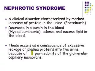
NephroticSyndrome The nephrotic syndrome is caused by renal diseases that increase the permeability across the glomerular filtration barrier Two issues are important in the pathogenesis of nephrotic syndrome: the mechanisms of glomerular injury and proteinuria.
Components of nephrotic syndrome • Proteinuria 3.5g/24 hr/1.73m2 • Hypoalbum. ( serum alb<3.5 g/d) • Hypercholesterolemia (>200mg/dl) • Peripheral edema ± anasarca
Tendencies of glomerulardiseasestomanifestNephroticFeatures • Minimal change glomerulopathy • Membranous GN • Diabetic GS • Amyloidosis • Focal segmental GS • Fibrilary GN
Common causes of NS • Primaryglomerulardisorders • Minimal ChangeDisease • FSGS • Membranous GN • Orthostatikorposturalproteinuria • Idiopatic MPGN • IgAnephropathy • Proliferatif GN
Common causes of NS • Secondary glomerular disorders Hereditary-familial: DM, Alport’s Syndrome, Sickle cell disease Autoimmun: SLE, Goodpasture’s syndrome, Wegener’s granulomatosis, PAN, RA Infectious: postinfectious glomerulonephritis, HIV FSGS Drug-induced: NSAIDs, Heroin,gold, mercury, Neoplastic: Hodgkin’s D., Lymphomas, leukemia, MM. miscellaneous: amyloidosis, preeclampsia-eclampsia, renovascular HT, intertitial nephritis, fever, exercise
Screening for Diabetic nephropathy When Normal Range • BP each visit • Urinary alb: Type 2: annually, beginning at diagnosis • Type 1: annually, 5 years post diagnosis • <130/80 • <30 mg/day • <20 mic/min • <30 mic/mg creatinine ADA 2004
Proteinuria podocyte • changestocapillaryendothelialcells, theglomerularbasementmembrane (GBM), orpodocytes, whichnormallyfilter serum protein selectivelybysizeandcharge. • Themechanism of damagetothesestructures is unknown in primaryglomerulardisease • Theresult is urinaryloss of macromolecularproteins, primarilyalbuminbut alsoopsonins, Ig’s, erythropoietin, transferrin, hormone-bindingproteins, andantithrombin III in conditionsthatcausenonselectiveproteinuria.
Pathophysiology of proteinuria • Glomerularretentionandleakage of protein molecules • Damage of glomeruler size andchargeselectivity Size:(molecularradius) <17 Åreadlypasstheglomerularfilter, >44 Å can not pass) • Albumin 36 Å Charge: glomerularcapillarywallfixnegativecharge • Normally 1500mg/24 hrProtein filtered, most is reabsorbed • <150 mg of protein excretedeachday in theurine
Types of proteinuria • 'Glomerular' proteinuria(more than about 1.5 g protein/24 h, mostly albumin) • 'Tubular' proteinuria(never excretion of more than 1.5 g/24 h ) • 'Overflow' proteinuria: immunoglobulin light chains in the urine • Benign proteinuria'Jogger's nephritis' , orthostatic proteinuria
Acute Complications of Nephrotic Syn. • peripheraledema, ascites, andeffusionsincreased risk forinfection (especiallycellulitisand, in 2 to 6%, spontaneousbacterialperitonitis); • anemia; abnormalthyroidfunction; • Thromboembolism (especiallyrenalveinthrombosisandpulmonaryembolism in upto 5% of childrenand 40% of adults). • Thromboembolismmaydevelop not onlybecause of urinaryloss of antithrombin III but alsobecause of increasedhepaticsynthesis of clottingfactors, plateletabnormalities, andhyperviscosityfromhypovolemia.
Hypercholesterolaemia is present in 90 per cent of patients with a urinary protein excretion of over 3 g/24 h • Hyperlipidemia in the NS is the result of both increased synthesis and decreased catabolism of lipoproteins.
Chronic complications of NS • malnutrition in children, • coronary artery disease in adults, • chronic renal failure, and bone disease. Malnutrition may mimic kwashiorkor, including brittle hair and nails, alopecia, and stunted growth. Coronary artery disease develops because NS causes hyperlipidemia, hypertension, and hypercoagulability.
Chronic complications of NS • Bone disease develops because of vitamin D deficiency and corticosteroid use. • hypothyroidism from loss of thyroid-binding globulin • proximal tubular dysfunction causing glucosuria, aminoaciduria, K depletion, phosphaturia, and renal tubular acidosis
Symptoms and Signs • anorexia, malaise, and frothy urine caused by high concentrations of protein. • Edema may cause dyspnea (pleural effusion or laryngeal edema), chest discomfort (pericardial effusion), • arthralgia (hydrarthrosis), • abdominal pain (ascites or, in children, mesenteric edema). • Edema may obscure signs of muscle wasting and cause parallel white lines in fingernail beds (Muehrcke's lines).
Diagnosis • suspected in patients with edema and proteinuria on urinalysis and confirmed by 24-h measurement of urinary protein. The cause may be suggested by history (eg, cancer); when the cause is unclear, serologic testing and renal biopsy are indicated. • Besides proteinuria, urinalysis may demonstrate RBCs and casts (hyaline, granular, fatty, waxy, RBC, or epithelial cell). • Lipiduria, the presence of free lipid or lipid within tubular cells (oval fat bodies), within casts (fatty casts), or as free globules
Evaluation for secondary causes • serum glucose or glycosylated hemoglobin (HbA1c), • antinuclear antibodies, • hepatitis B and C serologic tests,
Renal Bx • In adults, a renal biopsy is indicated to diagnose the underlying cause of idiopathic NS. • Idiopathic NS in children is most likely minimal change disease and is usually presumed without biopsy unless the patient fails to improve on a trial of corticosteroids.
NephriticSyndrome Defined by hematuria and RBC casts on microscopic examination of urinary sediment.
Components of ?Nephritic Syndrome • Often one or more elements of mild to moderate proteinuria, edema, hypertension, elevated serum creatinine, and oliguria are also present. • It has both primary and secondary causes. Diagnosis is based on history, physical examination, and sometimes renal biopsy. Treatment and prognosis vary by cause.
AsymptomaticHematuria • RecurrentGrossHematuria • Redbloodcell/ HP mic >3 cell • AsymptomaticHematuriaprevalence 5-10 % • TeaorColacoloredurine • Proteinuria< 1 g/24 hr • Serum Cr< 1.5 mg/dl
Tendencies of glomerulardiseasestomanifestNephriticFeatures • Proliferative GN(SLE) • Acute diffuse proliferative GN (Post Step. GN) • Crescentic GN(SLE, Wegener, Ig A) • Mesangioproliferative GN(Ig A)
Asymptomatic glomerular hematuria • No pathologic abnormality (30%) • Thin basement membrane (26%) • Ig A nephropathy (28%) • Alport syndrome ( rare ? Is it really rare in Turkey ?)
Alport's Syndrome • Hereditary nephritis is a genetically heterogenous disorder characterized by hematuria, impaired renal function, sensorineural deafness, and ocular abnormalities. • Cause is a gene mutation affecting type IV collagen. Symptoms and signs are those of nephritic syndrome with sensorineural deafness and, less commonly, those of ophthalmologic diseases. Diagnosis is by family history and urinalysis.
Alport's Syndrome • Because of X-linked transmission, women usually are asymptomatic and have little functional impairment. • Most men eventually develop renal symptoms and signs similar to those of acute nephritic syndrome and progress to renal insufficiency between ages 20 and 30.
Alport's Syndrome • Diagnosis is suggested by personal and family history and by findings of microscopic hematuria on urinalysis or recurrent episodes of gross hematuria, particularly if abnormalities of hearing or vision are present.
IgA nephropathy • It is the most common form of GN worldwide. • It occurs at all ages, with a peak onset in the teens and 20s; affects men 2 to 6 times more frequently than women; and is more common in whites and Asians than in blacks. • Prevalence estimates are 5% in the US, 10 to 20% in southern Europe and Australia, and 30 to 40% in Asia.
IgAnephropathy • Deposition of IgAimmunecomplexes in glomeruli, manifesting as slowlyprogressivehematuria, proteinuria, and, often, renalinsufficiency. • Diagnosis is based on urinalysisandrenalbiopsy. Prognosis is generallygood ?. Treatmentoptionsinclude ACE inhibitors, corticosteroids, andω-3 polyunsaturatedfattyacids.
PATHOGENESIS • Cause is unknown, • pathogenetic mechanisms, • increased IgA1 production, • defective IgA1 glycosylation causing increased binding to mesangial cells, • decreased IgA1 clearance, • a defective mucosal immune system, • overproduction of cytokines stimulating mesangial cell proliferation. • Familial clustering has also been observed, suggesting genetic factors at least in some cases.
Symptoms and Signs • persistent or recurrent macroscopic hematuria (90% of involved children) or • asymptomatic microscopic hematuria with mild proteinuria
Diagnosis • urinalysis and renal biopsy. • Urinalysis demonstrates microscopic hematuria, usually with dysmorphic RBCs and RBC casts. • Mild proteinuria (< 1 g/day) is typical and may occur without hematuria; nephrotic syndrome develops in ≤ 20%.
Ig A mesangial deposition granular deposition of IgA and C3 on immunofluorescent staining in an expanded mesangium with foci of segmental proliferative or necrotizing lesions.
PostinfectiousGlomerulonephritis • mostcommoncause of glomerulardisease in childrenbetween 5 and 15 yr; it is rare in children < 2 yrand in adults > 40 yr. • occurs after infection, usually with a nephritogenic strain of group A β-hemolytic streptococcus. • Diagnosis is suggested by history and urinalysis and confirmed by low complement.
Symptoms and signs • asymptomatic hematuria (in about 50%) and mild proteinuria • full-blown nephritis with microscopic or gross hematuria (cola-colored, brown, smoky, or frankly bloody), proteinuria, oliguria, edema, hypertension, and renal insufficiency. • Severe, late disease is a relatively uncommon cause of nephrotic syndrome. Flank pain may be attributable to stretching of the renal capsule. • Renal failure that causes fluid overload with heart failure and urgent or malignant hypertension and requires dialysis affects 1 to 2% of patients and may present as a pulmonary-renal syndrome with hematuria and hemoptysis
RapidlyProgressiveGlomerulonephritis (RPGN) • CrescenticGlomerulonephritis • RPGN causesmicroscopicglomerularcrescentformationwithprogressiontorenalfailurewithinweekstomonths. • Diagnosis is based on history, urinalysis, serologictests, andrenalbiopsy. • Treatment is withcorticosteroids, withorwithoutcyclophosphamide, andsometimesplasmapheresis.
Classification of RPGN Based on Immunofluorescence Microscopy
Anti-glomerular basement membrane (GBM) antibody disease (type 1 RPGN) • Thecombination of GNandalveolarhemorrhage in the presence of anti-GBM antibodies is calledGoodpasture'ssyndrome • autoimmune GN andaccountsfor 10% of RPGN cases. Itmayarisewhenrespiratoryexposures (eg, cigarettesmoke, viral URI) exposealveolarcapillarycollagen, triggeringformation of anticollagenantibodies. • Theanticollagenantibodiescross-reactwith GBM, fixingcomplementandtriggering a cell-mediatedinflammatoryresponse in thekidneysandlungs. Immunofluorescentstaining of renalbiopsytissuedemonstrateslinearIgGdeposits.