Download
1 / 13
330 likes | 1.55k Vues
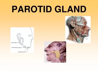
Parotid Salivary gland. It is the largest of all salivary glands. Shape: Irregular pyramid in shape. Position: It lies below the external acoustic meatus. It wedges between the ramus of the mandible and sternomastoid muscle.

E N D
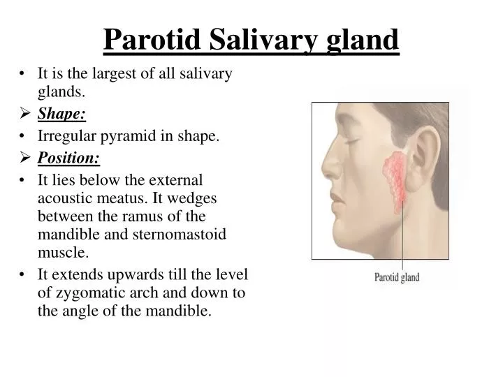
Parotid Salivary gland • It is the largest of all salivary glands. • Shape: • Irregular pyramid in shape. • Position: • It lies below the external acoustic meatus. It wedges between the ramus of the mandible and sternomastoid muscle. • It extends upwards till the level of zygomatic arch and down to the angle of the mandible.
The gland partially overlaps the masseter muscle. • A small semi-detached part of the gland lies between the zygomatic arch and the parotid duct which is called: accessory part of the parotid gland.
The gland has: • 2 ends; a superior end (surface), an inferior end which extends to to the level of angle of mandible. • 3 surfaces; superficial (lateral), anteromedial and posteromedial. • The gland is covered by a capsule derived from deep cervical fascia.
Main structures within the substance of the gland: • From superficial to deep, they are: • Facial nerve and branches. • Retromandibular vein and its two divisions, maxillary vein, superficial temporal vein.. • External carotid artery and its two terminal branches.
Facial nerve • It is the most superficial structure. • It enters the gland through its posteromedial surface. • It breaks within the gland into terminal branches that leave it through; upper end, lower end and anteromedial surface. • These terminal branches are 5; Temporal, zygomatic, buccal, marginal mandibular and cervical.
Other structures within the gland: • Auriculotemporal nerve. • Posterior auricular artery. • Preauricular lymph nodes.
Parotid duct: • The duct is about 5 cm in length. • It arises from anterior border of the gland. • It runs on the masseter muscle. • At the anterior border of masseter it runs medially to pierce the buccinator muscle and mucosa to open in the vestibule of the mouth.
Opening of parotid duct: It opens in the vestibule of the mouth opposite the upper second molar tooth. • Surface anatomy: The duct is represented by the middle 1/3 of the line between a point on the lower border of tragus of ear and a point midway between the ala of nose and red margin of the upper lip.
Blood supply: • Arterial: External carotid artery and its terminal branches within the gland. • Venous: Into the retromandibular vein. • Lymph drainage: Into Preauricular (parotid), superficial cervical and deep cervical lymph nodes.
Nerve supply: • Sensory supply: From Auriculotemporal nerve. • Sympathetic supply: From the plexus around external carotid artery. • Parasympathetic supply: Otic ganglion.
Parasympathetic supply: • From inferior salivary nucleus in brain stem. • To glosspharyneal nerve. • To its tympanic branch that breaks into tympanic plexus on the tympanic membrane. • The fibres then carried by lesser petrosal nerve to relay in otic ganglion. • Postganglionic fibers reach the gland via Auriculotemporal nerve.