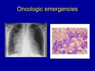
Oncologic Emergencies
Oncologic Emergencies. Javier Oesterheld MD Pediatric Hematology/Oncology. Oncologic Emergencies. Superior Vena Cava Syndrome (SVCS) Superior Mediastinal Syndrome (SMS) Spinal Cord Compression Metabolic Emergencies Infectious Complications Assorted Complications.


Oncologic Emergencies
E N D
Presentation Transcript
Oncologic Emergencies Javier Oesterheld MD Pediatric Hematology/Oncology
Oncologic Emergencies • Superior Vena Cava Syndrome (SVCS) • Superior Mediastinal Syndrome (SMS) • Spinal Cord Compression • Metabolic Emergencies • Infectious Complications • Assorted Complications
Pathophysiology • SVCS • The SVC is thin walled and surrounded by lymph nodes and thymus • Tumor or infection in the lymph nodes or thymus can compress the SVC • SMS • The trachea and main stem bronchi are compressible in children and tumor or infection in surrounding lymph nodes can lead to compression
SVCS and SMS • Etiologies • CV surgery • Shunting for hydrocephalus • Tumor • Most common primary cause • NHL, Hodgkin’s disease, ALL, GCT • Usually SCVS and SMS occur in the same patient
Symptoms • Cough, hoarseness, dyspnea, orthopnea, and chest pain • More advanced: anxiety, confusion, lethargy, headache, distorted vision, syncope • Symptoms are aggravated when patient is supine or flexed
Signs • Swelling, plethora and cyanosis of the face, neck and upper extremities • Suffusion and edema of the conjunctiva • Diaphoresis, wheezing, and stridor • Engorgement of the chest wall vessels • In children onset of symptoms are rapid
Evaluation • Management/diagnostic decisions difficult and controversial - - emergency treatment vs definitive Dx • Significant stridor, dyspnea usually not present unless airway cross-sectional area narrowed by >50% • Some authors recommend CT scan to evaluate tracheal compression prior to decisions regarding sedation/anesthesia
Evaluation • CXR • CT of Chest • AFP and beta HCG • Obtain diagnosis • The child may not tolerate GA – therefore the least invasive method of diagnosis should be used (i.e. CBC, BMA, Pleurocentesis)
Evaluation • Inability to tolerate supine position of grave significance with anterior mediastinal mass • May result from weight of tumor compressing not only airway, but great vessels and heart (especially RV outflow tract) • If can tolerate supine position, CT and PFT’s may help indicate which children will tolerate anesthesia • Shamberger, 1991 and 1995
Emergent Management • Keep child in sitting, left lateral decubitus position - - helps “lift” mass off airway and RVOT • IV access (lower extremities preferable due to SVC obstruction) • Face mask O2, non-invasive PEEP • Last resort is intubation!
Emergent Management • AVOID sedation for procedures unless anesthesiologist present and prepared for VERY difficult intubation • If impending respiratory failure and requires intubation . . . • Awake, bronchoscopic intubation ideal to maintain airway muscle tone, prevent worsened extrinsic compression • Ideally, have cardiopulmonary bypass available • AVOID neuromuscular blockade - - worsened obstruction even as low as carina
Therapy • Clot in SVC – Thrombolytic agent (TPA) • SMS – true emergency • Obtain Dx if possible • XRT • Often rapid improvement • There can be post irradiation tracheal swelling and deterioration • Chemotherapy • Steroids, cytoxan
Spinal Cord Compression • Occurs in 3-5% of children with cancer, often at diagnosis. • Can occur with any tumor type, but mostly with Leukemia, NHL neuroblastoma, sarcoma, and Hodgkin’s disease.
Spinal Cord Compression • Pathogenesis • Epidural compression most common in children • Metastatic tumor to vertebral body and secondary compression (Common in adults) is not common in children
Presentation Back Pain: suspect cord compression when . . . pain not relieved in supine position or back pain has a radicular component. • Weakness, sensory abnormalities, and paresis. • Paraplegia and quadriplegia can occur rapidly if there are neurologic abnormalities • Urinary and fecal incontinence.
Spinal Cord Compression • Signs and symptoms • Back pain in 80% • Abnormal neuro exam (esp. decreased motor strength)
Spinal Cord Compression • Evaluation • Plain films – 50% are normal • MRI • Therapy • Dexamethasone (initiate quickly if compression is a consideration) • Surgical decompression (unknown Primary) • Radiation therapy • Chemotherapy
Treatment • Symptoms of cord dysfunction: give dexamethasone and obtain MRI • Decompression: surgery, radiation, chemotherapy. • Surgery indicated if tumor type is not known or symptoms progress despite radiotherapy. • Chemotherapy is appropriate for patients with spinal cord compression due to lymphoma, leukemia, and neuroblastoma.
Metabolic Emergencies – Tumor Lysis Syndrome • TLS – is a consequence of rapid release of intracellular metabolites (uric acid, K+, phosphate) in quantities that exceed the excretory capacity of the kidneys • Usually occurs at diagnosis or within 5 days of starting therapy
Tumor Lysis Syndrome • Metabolic manifestations include • Increase UA, K+, phos • Secondary decreased calcium and cardiac arrhythmias • Renal failure • Most commonly seen in hematologic malignancies • Lymphoma (esp. Burkitt’s) • ALL • AML
Tumor Lysis Syndrome • Pathophysiology • Massive lyses of malignant cells • Worsened by inadequate renal function • Accelerated by treatment • Uric acid (calcium phosphate) precipitates in the kidney, causing worsening kidney function
Consequences of tumor lysis syndrome • Hyperkalemia weakness, dysrhythmias • Hyperphosphatemia hypocalcemia, renal failure • Hypocalcemia tetany, mental status changes, seizures • Hyperuricemia “uric acid nephropathy” = oliguria, renal failure
Tumor Lysis Syndrome • Labs to be obtained in at risk patients • CBC with diff • Electrolytes, BUN, creat, Ca++, Phos-, uric acid, LDH, and UA • CXR • If K+ increased, EKG looking for peaked T waves and/or QRS widening
Tumor Lysis Syndrome • Management • High Risk Patients • Burkitt’s lymphoma • Other NHL • Leukemia with increased WBC • Decreased urine output, increased creat • Metabolic abnormalities
Tumor Lysis Syndrome • Management (cont.) • Before cytotoxic chemotherapy started, metabolic stability should be achieved • For decreased urine output do renal US to R/O obstructive uropathy • Hydration • 3000cc/msq/d – NO K+ • Maintain SG < 1.010 and /or 4cc/kg/hr
Tumor Lysis Syndrome • Management (cont.) • Alkalinization • > 50meq/L NaHCO3 – adjust to keep ph 7.0 – 7.5 • Diamox 150 – 1000mg/msq/d divided q6 • Avoid ph > 7.5 due to crystallization of CaPhos • Allopurinol – 300 mg/msq/day po or IV • Rasburicase
Tumor Lysis Syndrome • Hypoxanthine* Xanthine* Uric Acid-> Allantoin • *Xanthine Oxidase involved in reaction • Inhibited by Allopurinol • Urate Oxidase involved in reaction Uric Acid -> Allantoin • Allantoin more soluble than UA or Xanthine • Recombinant urate oxidase (Rasburicase) used for severe TLS
Tumor Lysis Syndrome • Management • Inadequate urine output • Lasix 0.5 – 1 mg/kg q4 hr or 1mg/kg/hr CI • Mannitol 15 gm/msq q6 hr IV over 30 min • Hyperkalemia - > 6.0 – 6.5 meq/L with EKG changes • Kayexalate: 1 gm/kg po or pr with 50% sorbitol • Insulin (Reg 0.1 u/kg) in 2 cc/kg 25% DEX IV • Initiate steps to begin dialysis
Tumor Lysis Syndrome • Management • Hyperphosphatemia • Aluminum hydroxide (Amphogel) : 50 – 150 mg/kg/d with meals • Hypocalcemia, symptomatic • 10% calcium gluconate 0.5 – 1 mg/kg IV • Dialysis
5-20% of children with new Dx of leukemia have WBC count > 100,000/mm3 These patients at risk of severe complications from hyperviscosity of blood Hyperleucocytosis
Hyperleukocytosis - Complications • Blasts interact with endothelium to form aggregates, thrombi in microcirculation • Most problems in CNS and pulmonary circulation • Complications more common with AML than ALL • Myeloblasts and monoblasts larger, less deformible, “stickier”
Pulmonary leukostasis • Sx: dyspnea, tachypnea, hypoxemia, acidosis, cor pulmonale • CXR: diffuse interstitial infiltrates
CNS Manifestations • Headache, mental status changes, seizures, coma in spectrum of Sx • High risk of intracranial hemorrhage, especially with AML and thrombocytopenia
Therapy for hyperleukocytosis • Decrease blood viscosity (directly related to morbidity) • Hydration • AVOID use of diuretics • AVOID PRBC transfusion (Hb goal < 10 gm/dL for viscosity) • Transfuse platelets to keep > 20,000/mm3 and treat coagulopathy (common with AML) to decrease risk of intracranial hemorrhage
Therapy for hyperleukocytosis • Urine alkalinization, hydration, etc as with tumor lysis syndrome • Consider leukapheresis or exchange transfusion • PICU supportive care - mechanical ventilation, hemodynamic support, etc
Same diagnostic criteria as other pts: Fever/hypothermia Tachycardia Tachypnea Hypoperfusion Acidosis Hypotension (SCCM/ACCP Consensus Conference, 1992) Common etiologies: Gram + cocci -hemolytic Strep Staph. Epi Staph aureus Gram - rods Pseudomonas Enterobacter E. coli Fungi Candida spp Viruses Sepsis in Pediatric Cancer Patients
Infectious Complications • Risk Factors • Alterations of cellular and humoral immunity • Disruption of natural barriers • Colonization with more pathogenic organisms • Abnormalities of lymphocytic, spleen, and RES • Decreased PMN
Infectious Complications • Definition • Fever • >38.5 or 38.0 twice within 12 hour period • Neutropenia • ANC < 500 or less than 1000 and predicted to be less than 500 within 24 – 48 hours
Infectious Complications • Fever – Nonneutropenic/No CV line • PE – non toxic child • No source of infection or defined non-life threatening source • Obtain blood and urine cultures and other appropriate cultures • Treat identified infection as appropriate • Close F/U, no hospitalization or IV antibiotics necessary
Infectious Complications • Fever – Non-neutropenic/indwelling CV line • PE - non-toxic child, no evidence of likely systemic infection • Blood cultures through all lumens • Urine culture and other cultures as appropriate • Hospitalization and IV antibiotics • Coverage for both gram + and gram – organisms • Continue antibiotics for 48 – 72 hours and then D/C if cultures negative • Observation with or w/o IV antibiotics also acceptable
Infectious Complications • Fever/Neutropenic patient • Thorough history and PE • Especially sites commonly a source of infection: skin, lungs, perioral, and perirectal area • Subtle signs of inflammation should be considered signs of infection • Blood and urine cultures and other cultures as appropriate • CXR, LP, wound cultures as appropriate • Hospitalize and treat with IV antibiotics
Infectious Complications • Consideration for antibiotic choice • < 50% will have defined site of infection • 85 – 90% of identified pathogens associated with new fever in neutropenic patients are bacterial • Gram (+) and Gram (–) bacteria involved
Infectious Complications • Treatment considerations • Documented infections are treated 10 – 14 days • Patients can be divided into 2 risk groups • Low risk group – ANC expected to recover within 7 days and there is no co-morbidity (decreased BP, respiratory compromise, altered MS) • High risk group – ANC expected to be low for > 7 days
Infectious Complications • Treatment Duration • Low risk group – D/C antibiotics when ANC recovering (increasing monos/platelets/WBC) and blood cultures negative and afebrile for 24 hours • High risk group • Patients who become afebrile within 7 days but remain neutropenic should have antibiotics continued until ANC recovering or 14 days
Infectious Complications • High risk group who remains febrile >3-4 days • Continue antibiotics, but add antifungal therapy • If Abx were stopped in febrile neutropenic patients at day 7, 56% developed symptoms of infection with 3 days, 36 % presented with hypotension • If Abx continued 31 % developed invasive fungal
Infectious Complications • Rationale of treatment for high risk group • In patients who became afebrile but remained neutropenic and had antibiotics stopped at day 7, 41% became febrile within 3 days – 0% became febrile if antibiotics continued • Patients who were afebrile but neutropenic and had antibiotics stopped at day 14 had the same incidence of recurrent fevers as those who continued on antibiotics past 14 days – 30%