Figure 22 Section 2
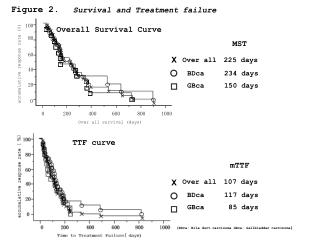
Figure 22 Section 2. Steps in the Process of Digestion. In the oral cavity, saliva dissolves some organic nutrients, and mechanical processing with the teeth and tongue disrupts the physical structure of the material and provides access for digestive enzymes. Those enzymes begin
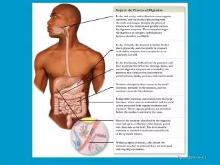

Figure 22 Section 2
E N D
Presentation Transcript
Figure 22 Section 2 Steps in the Process of Digestion In the oral cavity, saliva dissolves some organic nutrients, and mechanical processing with the teeth and tongue disrupts the physical structure of the material and provides access for digestive enzymes. Those enzymes begin the digestion of complex carbohydrates (polysaccharides) and lipids. In the stomach, the material is further broken down physically and chemically by stomach acid and by enzymes that can operate at an extremely low pH. In the duodenum, buffers from the pancreas and liver moderate the pH of the arriving chyme, and various digestive enzymes are secreted by the pancreas that catalyze the catabolism of carbohydrates, lipids, proteins, and nucleic acids. Nutrient absorption then occurs in the small intestine, primarily in the jejunum, and the nutrients enter the bloodstream. Indigestible materials and wastes enter the large intestine, where water is reabsorbed and bacterial action generates both organic nutrients and vitamins. These organic products are absorbed before the residue is ejected at the anus. Most of the nutrients absorbed by the digestive tract end up in a tributary of the hepatic portal vein that ends at the liver. The liver absorbs nutrients as needed to maintain normal levels in the systemic circuit. Within peripheral tissues, cells absorb the nutrients needed to maintain their nutrient pool and ongoing operations.
Digestion, Absorption, Transport • Digestion • Breakdown of food molecules for absorption into circulation • Mechanical: Breaks large food particles to small • Chemical: Breaking of covalent bonds by digestive enzymes • Absorption and transport • Molecules are moved out of digestive tract and into circulation for distribution throughout body
Nervous regulation Involves enteric nervous system Types of neurons: sensory, motor, interneurons Coordinates peristalsis and regulates local reflexes Chemical regulation Production of hormones Gastrin, secretin Production of paracrine chemicals Histamine Help local reflexes in ENS control digestive environments as pH levels Digestive System Regulation
Digestive System Anatomy • Digestive tract • Alimentary tract or canal • GI tract • Accessory organs • Primarily glands • Regions • Mouth or oral cavity • Pharynx • Esophagus • Stomach • Small intestine • Large intestine • Anus
Peritoneum and Mesenteries • Peritoneum • Visceral: Covers organs • Parietal: Covers interior surface of body wall • Retroperitoneal: Behind peritoneum as kidneys, pancreas, duodenum • Mesenteries • Routes which vessels and nerves pass from body wall to organs • Greater omentum • Lesser omentum
Oral Cavity • Mouth or oral cavity • Vestibule: Space between lips or cheeks and alveolar processes • Oral cavity proper • Lips (labia) and cheeks • Palate: Oral cavity roof • Hard and soft • Palatine tonsils • Tongue: Involved in speech, taste, mastication, swallowing
Teeth • Two sets • Primary, deciduous, or milk: Childhood • Permanent or secondary: Adult (32) • Types • Incisors, canine, premolar and molars
Salivary Glands • Produce saliva • Prevents bacterial infection • Lubrication • Contains salivary amylase • Breaks down starch • Three pairs • Parotid: Largest • Submandibular • Sublingual: Smallest
Deglutition (Swallowing) • Three phases • Voluntary • Bolus of food moved by tongue from oral cavity to pharynx • Pharyngeal Reflex: Upper esophageal sphincter relaxes, elevated pharynx opens the esophagus, food pushed into esophagus • Esophageal • Reflex: Epiglottis is tipped posteriorly, larynx elevated to prevent food from passing into larynx
The process of peristalsis Bolus of food arrives in digestive system. Food bolus Toward anus Longitudinal muscle Circular muscle Circular muscles contract behind bolus. Longitudinal muscles ahead of bolus contract. Contraction in circular muscle layer forces bolus forward.
Stomach Anatomy: • Openings • Gastroesophageal: To esophagus • Pyloric: To duodenum • Regions • Cardiac • Fundus • Body • Pyloric
Stomach Anatomy cont. • Rugae: Folds in stomach when empty • Gastric pits: Openings for gastric glands • Contain cells • Surface mucous: Mucus • Mucous neck: Mucus • Parietal: Hydrochloric acid and intrinsic factor • Chief: Pepsinogen • Endocrine: Regulatory hormones
The structure of the wall of the stomach Layers of the Stomach Wall Mucosa Consists of a simple columnar epithelium that produces an alkaline carpet of mucus that covers the interior surfaces of the stomach and protects epithelial cells against the acid and enzymes in the gastric lumen Lymphatic vessel Lamina propria Muscularis mucosae Artery and vein Submucosa Muscularis Externa Oblique muscle Circular muscle Myenteric plexus Longitudinal muscle Serosa
Figure 21.9 2 The structure of gastric pits and gastric glands Lamina propria Mucous epithelial cells Gastric pit Neck Cells of Gastric Glands Parietal cells (secrete HCl and intrinsic factor) G cells (produce a variety of hormones) Gastric glands Chief cells (secrete pepsinogen)
Figure 21.9 3 The secretory activities of parietal cells Hydrogen ions (H+) are generated inside a parietal cell as the enzyme carbonic anhydrase converts CO2 and H2O to carbonic acid (H2CO3), which then dissociates. KEY Parietal cell Diffusion Carbonic anhydrase Carrier-mediated transport The hydrogen ions are actively transported into the lumen of the gastric gland. A countertransport mechanism ejects the bicarbonate ions into the interstitial fluid and imports chloride ions into the cell. Active transport Countertransport Interstitial fluid The chloride ions then diffuse across the cell and exit through open chloride channels into the lumen of the gastric gland. Lumen of gastric gland To bloodstream
Ingested food The pattern of hormone release and the effects of those hormones within the digestive system Hormone Action KEY Food in stomach inhibits stimulates Acid production by parietal cells Gastrin Stimulation of gastric motility; mixing waves increase in intensity Release of insulin from pancreas GIP Release of pancreatic enzymes and buffers Chyme in duodenum Secretin and CCK Bile secretion and ejection of bile from gallbladder facilitates Dilation of intestinal capillaries VIP facilitates NUTRIENT UTILIZATION BY ALL TISSUES Material arrives in jejunum Nutrient absorption
Figure 21.13 2 The two central reflexes triggered by the stimulation of stretch receptors in the stomach wall Central Gastric Reflexes Gastroenteric reflex: stimulates motility and secretion along the entire small intestine Gastroileal reflex: triggers the opening of the ileocecal valve, allowing materials to pass from the small intestine into the large intestine Ileocecal valve
Small Intestine • Site of greatest amount of digestion and absorption • Divisions • Duodenum • Jejunum • Ileum: Peyer’s patches or lymph nodules • Modifications • Circular folds or plicae circulares, villi, lacteal, microvilli • Cells of mucosa • Absorptive, goblet, granular, endocrine
Figure 21.11 2 The characteristic features of each of the three segments of the small intestine Jejunum Serosa Duodenum Muscularis externa Duodenal glands Plicae circulares Submucosa Mucosa Muscularis mucosae Villi Ileum Aggregated lymphoid nodules
Small Intestine Secretions • Mucus • Protects against digestive enzymes and stomach acids • Digestive enzymes • Disaccharidases: Break down disaccharides to monosaccharides • Peptidases: Hydrolyze peptide bonds • Nucleases: Break down nucleic acids • Duodenal glands • Stimulated by vagus nerve, secretin, chemical or tactile irritation of duodenal mucosa
Mixing: Segmental contraction that occurs in small intestine Involves contraction of circular muscles only
Figure 21.10 Intestinal adaptations for absorbing nutrients A photomicrograph showing the brush border of an intestinal villus The structure of an intestinal villus Capillaries The complex internal structure of an intestinal villus Plica circulares Villi Mucous cells A plica circulares and villi in the small intestinal wall Lacteal Brush border Tip of villus LM x 250 A diagrammatic sectional view of the intestinal wall showing features common to all segments of the small intestine Columnar epithelial cell Villi Submucosal artery and vein Lacteal (lymphatic capillary) Mucous cell Lacteal Nerve Layers of the Small Intestine Intestinal gland Capillary network Mucosa Arteriole Lymphatic vessel Lamina propria Muscularis mucosae Venule Lymphoid nodule Submucosa Submucosal plexus Muscles that move the villi back and forth to expose the epithelial surfaces to the intestinal contents Circular layer of smooth muscle Muscularis externa Myenteric plexus Serosa Longitudinal layer of smooth muscle Muscularis mucosae Lymphatic vessel
Figure 21.10 1 – 3 Intestinal adaptations for absorbing nutrients Plica circulares Villi A plica circulares and villi in the small intestinal wall A diagrammatic sectional view of the intestinal wall showing features common to all segments of the small intestine Lacteal (lymphatic capillary) Submucosal artery and vein Villi Layers of the Small Intestine Intestinal gland Mucosa Muscularis mucosae Lymphoid nodule Submucosa Submucosal plexus Circular layer of smooth muscle Muscularis externa Myenteric plexus Serosa Longitudinal layer of smooth muscle Lymphatic vessel
Figure 21.10 2 Plica circulares Villi A plica circulares and villi in the small intestinal wall
Figure 21.10 3 A diagrammatic sectional view of the intestinal wall showing features common to all segments of the small intestine Lacteal (lymphatic capillary) Submucosal artery and vein Villi Layers of the Small Intestine Intestinal gland Mucosa Muscularis mucosae Lymphoid nodule Submucosa Submucosal plexus Circular layer of smooth muscle Muscularis externa Myenteric plexus Serosa Longitudinal layer of smooth muscle Lymphatic vessel
Figure 21.10 4 – 5 A photomicrograph showing the brush border of an intestinal villus The structure of an intestinal villus Capillaries The complex internal structure of an intestinal villus Mucous cells Lacteal Brush border Tip of villus LM x 250 Columnar epithelial cell Mucous cell Lacteal Nerve Capillary network Arteriole Lymphatic vessel Lamina propria Venule Muscles that move the villi back and forth to expose the epithelial surfaces to the intestinal contents Muscularis mucosae
Accessory Glands and Structures • Liver • Gall bladder • Exocrine Pancreas • Pancreatic duct • Hepatic Portal System
Anatomy Endocrine Pancreatic islets produce insulin and glucagon Exocrine Acini produce digestive enzymes Regions: Head, body, tail Secretions Pancreatic juice (exocrine) Trypsin Chymotrypsin Carboxypeptidase Pancreatic amylase Pancreatic lipases Enzymes that reduce DNA and ribonucleic acid Pancreas
Exocrine Pancreas – Enzymes • Trypsinogen • Chymotrysinogen • Carboxypeptidases • Pro-elastase • Phospholipase • pancreatic lipase • Pancreatic amylase • Enzymes that reduce DNA and ribonucleic acid
Gallbladder • Bile is stored and concentrated • Stimulated by cholecystokinin and vegal stimulation • Dumps into small intestine • Production of gallstones possible • Drastic dieting with rapid weight loss
Liver • Lobes • Major: Left and right • Minor: Caudate and quadrate • Ducts • Common hepatic • Cystic • From gallbladder • Common bile • Joins pancreatic duct at hepatopancreatic ampulla
Functions of the Liver • Bile production • Salts emulsify fats, contain pigments as bilirubin • Storage • Glycogen, fat, vitamins, copper and iron • Nutrient interconversion • Detoxification • Hepatocytes remove ammonia and convert to urea • Phagocytosis • Kupffer cells phagocytize worn-out and dying red and white blood cells, some bacteria • Synthesis • Albumins, fibrinogen, globulins, heparin, clotting factors