Download

1 / 50
530 likes | 699 Vues
Learn about pemphigus vulgaris, an autoimmune blistering disease that mainly affects middle-aged and elderly individuals. Discover its clinical features, diagnosis methods, and treatment options involving steroids and immunosuppressants.

E N D
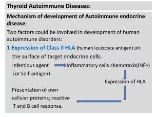
http:www.srrsh.com Autoimmune blistering diseases Pemphigus Pemphigoid Dermatitis herpetiformis (Duhring) Dr. Kejian Zhu Sir Run Run Shaw Hospital
Blister---classification • Intraepithelial vesicles: Acantholytic vesicles: the break down of specialized attachments (desmosomes) Nonacantholytic vesicles: the death or the rupture of the group of cells, usually seen in viral infections • Subepithelial vesicles:
Autoimmune blistering diseases • Diseases with intraepidermal blistering (pemphigus-group) Pemphigus vulgaris Pemphigus vegetans Pemphigus foliaceus Pemphigus erythematosus Paraneoplastic pemphigus Drug-induced pemphigus Neonatal pemphigus Intercellular IgA dermatosis Brazilian pemphigus
Autoimmune blistering diseases • Diseases with subepidermal blistering (pemphigoid group) Bullous pemphigoid (BP) Herpes gestationis Cicatricial pemphigoid Epidermolysis bullosa acquisita Dermatitis herpetiformis (Duhring) Linear IgA bullous dermatosis (LAD) Bullous SLE
Diseases with intraepidermal blistering (pemphigus-group) Pemphigus vulgaris Pemphigus vegetans Pemphigus foliaceus Pemphigus erythematosus
Outline • Middle-aged and elderly people are most commonly affected • Flaccid blisters • Two main groups: vulgaris & foliaceus • Nikolsky’s sign and Tzanck test are positive • Autoimmune diseases • Acantholytic intraepidermal blistering is produced by autoantibodies against desmoglein • Anti-desmoglein antibodies are detected by ELISA • In vivo IgG deposition and IgG antibodies are observed by immunofluorescence • Oral steroids & immunosuppressants are mainly administered.
Adhesion of keratinocytes Keratinocytes are firmly adhered by desmosomes. Transmembrane adhesion molecules in the cadherin superfamily, such as desmoglein 1 (Dsg 1), Dsg3, and desmocolin cadherin (DC), are important to intercellular adhesion. In pemphigus, autoantibodies are produced against Dsg1 and Dsg3, some of whose molecular functions are disturbed. This causes acantholysis.
Acantholysis • Acantholysis: dissociation of intercellular connections in the epidermis. As dissociation progresses, epidermal cleavage and blistering occurs. • Acantholytic cells: deformed keratinocytes become spherical from loss of intercellular connection within the blisters.
Tzanck test • Tzanck test is a kind of cytological diagnosis method. • It is induced by applying a slide glass to the bottom of a broken blister and staining the adhered cellular components in Giemsa for observation under a light microscope. • Tzanck cells are acantholytic cells observed in pemphigus. • Tzanck cells can also be observed in blisters of herpes simplex and herpes zoster, ballooning cells produced by viral infection.
Nikolsky’s sign • Nikolsky's sign: blistering or exfoliation of the skin’s outmost layer produced by slight rubbing of the normal-looking skin • It is positive in pemphigus, epidermolysis bullosa, staphylococcal scalded-skin syndrome (SSSS), and toxic epidermal necrolysis (TEN). • Nikolsky's sign is useful in differentiating between pemphigus vulgaris (where it is present or positive) and bullous pemphigoid (where it is absent)
Pemphigus vulgarisPemphigus vegetansPemphigus foliaceusPemphigus erythematosus
Outline • The most common variety of pemphigus • The disease most frequently occurs in the middle-aged and elderly. • The disease is caused by autoantibodies against desmoglein 3, which is a desmosomal adhesion factor in keratinocytes. • Acantholytic blisters form immediately above epidermal basal cells。 • It tends to manifest as oral enanthema. • Nikolsky’s sign is positive. • Oral steroids and immunosuppressants are the first-line treatment.
Clinical features • Most frequently affects the middle-aged and elderly • Erosions and ulcers develop acutely in the oral mucosa in 70-80% of cases • Blisters of various sizes occur on normal skin, easily rupture to form erosions and crusts • Painful, esp. when touched • Anywhere on the body, esp. at sites of pressure and friction (back, buttocks and feet) • Nikolsky’s sign • When widespread, electrolyte abnormalities due to loss of body fluid or hypoprotein • Be fatal when there is secondary infection
Workup • Pathology: acantholysis, intraepidermal blistering, leaving one basal layer at the bottom • Immunofluorescence: intercellular in vivo IgG deposition • ELISA: anti-Dsg3 IgG Ab, sometimes also anti-Dsg1 IgG Ab
Diagnosis • Clinical features • Pathology • Immunofluorescence • ELISA • DDx: bullous pemphigoid, impetigo, bullous drug eruption, dermatitis herpetiformis, erythema multiforme, Stevens-Johnson syndrome, etc.
Treatment • Systemic application of steroids is the first-line treatment (0.5-1.0mg/kg/d). • Taper off to a maintenance dose or until it can be discontinued. • Immunosuppressants (mycophenolate mofetil, CTX, AZT, MTX, cyclosporine) may be used. • In intractable cases, plasma exchange therapy and IVIG can be performed. • Antibiotics, fluid transfusion, nutrition management are conducted supplementarily.
Pemphigus vulgarisPemphigus vegetansPemphigus foliaceusPemphigus erythematosus
Clinical features • A subtype of pemphigus vulgaris, the most uncommon variety of pemphigus • Characterized by the formation of vesicles and erosions that do not re-epithelialize but gradually proliferate and elevate • Frequently occurs on areas of friction (axillary fossa, umbilical fossa, periphery of the oculonasal and perioral regions) and exposure (face, neck, scalp) • Oral mucosa is often involved • Strong odor
Workup • Pathology: suprabasal cell acantholysis, downward proliferation of rete ridges • Immunofluoscence: intercellular deposition of IgG and C3 • Culture: bacteria and/or candida
Diagnosis • Vesicles and erosions • Areas of friction, exposure • Proliferate and elevate • Suprabasal cell acantholysis • DDx: chronic pyoderma and fungal granuloma, Hailey-Hailey disease, condyloma acuminatum, etc.
Treatment • The same as for pemphigus vulgaris • Treat local infections Consider topical and systemic antibiotics Consider antifungal agents for candida • Surgical excision of large vegetative growths • Better prognosis than pemphigus vulgaris
Pemphigus vulgarisPemphigus vegetansPemphigus foliaceusPemphigus erythematosus
Outline • Autoantibodies are produced exclusively against Dsg 1 • Acantholysis and blistering are seen in the superficial epidermis (in the granular cell layer) • Fragile blisters, scaling and erosion, accompanied by crusts • Lesions are not/occasionally produced in the mucosa • Examinations and treatments are the same as for pemphigus vulgaris. The steroid dosage is usually less than for pemphigus vulgaris
Clinical features • Most commonly affects the middle-aged and elderly • Extremely fragile flaccid vesicles • Some of the blisters dry to become leafy and to exfoliate successively • The face, head, back and chest are most commonly affected • When spreads over the whole body, it resembles exfoliative erythroderma • Mucosa is not or occasionally involved • Nikolsky’s sign is positive
Workup • Pathology: acantholytic blistering is found in the epidermal upper layer. • Immunofluorescence: intercellular in vivo IgG deposition is observed • ELISA: anti-desmoglein 1 antibodies
Diagnosis • Clinical features • Pathology • Immunofluorescence • ELISA • DDx: pemphigus vulgaris, pemphigus erythematosus, drug-induced bullous disease, paraneoplastic pemphigus, etc.
Treatment • The same as for pemphigus vulgaris • Oral steroid dosage may be less than that for pemphigus vulgaris • In limited involvement cases, topical steroid are sufficient
Pemphigus vulgarisPemphigus vegetansPemphigus foliaceusPemphigus erythematosus
Clinical features • A subtype of pemphigus foliaceus • Occurs most commonly in the middle-aged and elderly • Frequently affects the seborrheic zones (head, face, chest and back) • The mucosa is not involved • Involvement of SLE is seen in some cases
Workup • Pathology: intraepidermal superficial bullae within the granular layer or just below it. Acantholysis may occur in the blister floor or roof. • Immunofluorescence: linear deposits of IgG and C3 in the intercellular space of the epidermis • May have lab abnormalities of SLE
Diagnosis • Clinical features • Pathology • Immunofluorescence • DDx: paraneoplastic pemphigus, seborrheic dermatitis, lupus erythematosus, pemphigus foliaceus, etc.
Treatment • The same as for pemphigus foliaceus
Diseases with subepidermal blstering (pemphigoid group) Bullous pemphigoid (BP) Dermatitis herpetiformis (Duhring)
Outline • Subepidermal blistering occurs as a result of autoantibody action against epidermal basement membrane structural proteins • Blisters are tense and do not rupture easily • Divided into pemphigoid, linear IgA bullous dermatosis, epidermolysis bullosa acquisita, dermatitis herpetiformis, herpes gestations, etc. • Immunofluorescence is useful for diagnosis • Steroids and dapsone are applied
Basement membrane zone • the area corresponding to the dermo-epidermal junction stains with periodic acid-Schiff consists of the basal cell plasma membrane the lamina lucida the basal lamina the sub-basal lamina fibrous components • acts as a mechanical barrier and penetration of substance between dermis and epidermis
pathogenesis • Autoantibodies are produced against hemidesmosome, type XVII collagen (BP180) and BP230 in the epidermal basement membranes, which leads to blistering. • Autoantibodies against BP180 play a major role.
Outline • Autoantibodies against hemidesmosomes • The major pathogenic antigen is type XVII collagen (BP180). • The roof of the blister has the full thickness of the epidermis • Elderly people account for the majority of cases • Characterized by subepidermal blisters • Blisters do not rupture easily • Oral steroids are effective
Clinical features • The elderly are more commonly affected • Multiple relatively large and severe tense blisters form immediately • Often accompanied by edematous erythema • Much less invasively to the mucous membranes (20% involved) • The general condition is favourable • May be complicated by malignant tumors
Workup • Pathology: subepidermal blistering, accompanied by eosinophilic infiltration • Immunofluorescence: linear IgG and C3 deposition in the basement membranes • ELISA: autoantibodies against type XVII collagen (BP180) proteins • High IgE values and elevated levels of eosinophils in peripheral blood
Diagnosis • Clinical features • Pathology • Immunofluorescence • ELISA • DDx: drug-induced bullous disorders, epidermolysis bullosa, epidermolysis bullosa acquisita, erythema multiforme, dermatitis herpetifomis, linear IgA dermatosis, etc.
Treatment • Oral steroids (0.5mg/kg/d) • Gradually reduced • Combination therapy of Immunosuppressants (CTX), DDS, tetracyclines and nicotinic-acid amide are also useful • Avoid secondary infections • Nutrition management is important for elderly • Topical steroid application may be sufficient in mild cases • Plasma exchange therapy and IVIG may also be used in severe cases
Outline • Characterized by extremely intense itching and irritation, chronically recurrent erythema and vesicles • Vesicles tend to form circular patterns • Common in Caucasians, rare in Asians • Granular IgA deposition in the dermal papillary • Gluten-induced enteropathy develops as a complication • Oral dapsone is effective
Gluten composed of the sticky, storage proteins found in wheat exist conjoined with starch in the same grass-related grains, notably wheat, rye and barley
Pathogenesis • IgA antibodies against tissue transglutaminase • The granular IgA deposition in the skin is an immuno-complex
Clinical features • Extremely intense itching • Erythema and urticarial lesions • Vesicles in a ring-shaped pattern • Scratch and resulted crusts • Heal with abnormal pigmentation or depigmentation • Appear symmetrically on the entire body, esp. on the elbows, knees and buttocks • Gluten-induce enteropathy is found in more than 90% of cases
Workup • Pathology: subepidermal blistering, micro-abscesses of neutrophils in dermal papillary • Immunofluorescence: granular IgA deposition in the dermal papillary
Diagnosis • Clinical features • Pathology • Immunofluorescence • DDx: linear IgA bullous dermatosis, bullous pemphigoid, herpes gestationis, erythema multiforme, ect.
Treatment • Dapsone is effective • Gluten-free diet • antihistamines