Lactation Physiology (part 1)
570 likes | 930 Vues
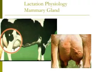
فیزیولوژی تولید و ترشح شیر. Lactation Physiology (part 1). By: A. Riasi (PhD in Animal Nutrition & Physiology). What is a mammary gland?. Serves a reproductive function; nourishment of the neonate.

Lactation Physiology (part 1)
E N D
Presentation Transcript
فیزیولوژی تولید و ترشح شیر Lactation Physiology(part 1) By: A. Riasi (PhD in Animal Nutrition & Physiology)
What is a mammary gland? • Serves a reproductive function; nourishment of the neonate. • The mammary gland is one of a few tissues in mammals, which can repeatedly undergo growth, functional differentiation, and regression • Relies on same endocrine (hormonal) support for development and function. • Example: gonadal steroids, prolactin, etc.
What is the difference between the animal udder? • Cow: Four glands and four teats • Sheep and goats: Two glands and two teats • Sow: 12-14 teats and two glands per teat. • Mare: Four glands and only two teats.
The udder is a complex system • A supportive system. • A secretory system composed of epithelial cells. • A duct system for storage and conveyance of milk. • Blood, lymph, and nerve systems.
The weight of empty cows udder is about 12-30 kg. • The udder weight is affected by: • Age • Stage of lactation • Amount of milk in the udder • Inherited differences among cows
There are seven tissues that provide support for the udder: • Skin covering the gland is only of very minor support. • Superficial fascia or Areolar subcutaneous tissue • Coarse areolar or cordlike tissue • Subpelvic tendon • Superficial layers of lateral suspensory ligament • Deep lateral suspensory ligament • Median Suspensory Ligament
An illustrated view of the ligaments that permit udder suspension (Courtesy of Iowa State University)
Interior anatomy of the Mammary Gland • The interior structure of mammary gland: • Connective tissue • Ductular system • Secretory tissue
Secretory tissue • A lactating secretory cell is the basic unit of milk synthesis • Milk precursors are taken from the blood into the cell • The secretory cell have two kind of junctions with neighbor cells: • Tight junction around the apical portion • Gap junction in lateral portion
Major component of a secretory epithelial cell Apical membrane Golgi apparatus Secretory vesicles Tight junction Gap junction Endoplasmic Reticulum Nucleus Lysosomes Basal and lateral membranes Cytoplasm Basement membrane
Precursors of Milk • Precursors of milk come from the bloodstream and the primary substrates extracted from blood include: • Glucose • Amino acids • Fatty acids • Minerals • Acetate * • βHB *
Precursors of Milk • Several materials in milk come unchanged from the blood: • Minerals • Hormones • Immunoglobulins
Synthesis of milk proteins • There are several specific systems for amino acids are absorption through the basal membrane. • Inside the cell, amino acids are covalently bound together to form proteins at the polysomes (Poly-ribosomes). • Proteins sythesized at RER include: • Casein • β-lactoglobulin, and α-lactalbumin • Membrane bound proteins
Synthesis of milk proteins • Synthesized proteins are transferred the golgi apparatues (GA). • Casein is secreted as micelle, which is formed in the GA from: • Casein molecues • Calcium • Phosphorus
Synthesis of milk lactose • Glucose enters the cells via the basolateral membrane via specific transport system. • Some glucose is converted to galactose in the cell. • Both glucose and galactose enter the GA and react resulting in the formation of lactose.
Synthesis of milk fat • In ruminant, acetate and β-hydroxybutyrate are important precursors of fatty acids (FA) synthesis in mammary cells. • Preformed FA, glycerol and monoacylglyceride are absorbed at the basolateral membrane. • Milk fat triglycerides are synthesized on the smooth endoplasmic reticulum and form small droplet.
Synthesis of milk fat • Under certain circumstances, fat droplets fuse with each other to form cytoplasmic vacuoles. • The protein coat on the milk fat globule membrane comprises: • Mainly butyrophilin (BTN) * • Xanthine oxidoreductase (XDH) * • Adipophilin (ADPH) • Mucin 1 • CD36 • Periodic acid/Schiff • PAS III, and FABP
Pathways for milk fat globule transit and secretion from mammary epithelial cells
Synthesis of milk fat • The properties of milk fat: • Milk fat composed of different fatty acids: • Short chains (4-8 C) • Medium chains (10-14 C) • Long chains (≥16 C) • More than 95% of milk fat is TAG • Around 70% of FA by weight in milk fat are saturated • Approximately 25% of the milk FA are monounsaturated • PUFA only account for a small portion of total FA in milk
Synthesis of milk fat • There are two sources of FA for milk fat synthesis: • The de novo FA synthesis in mammary epithelial cells • Short chain (4-8 C) • Medium chain (10-14 C) • About 50% of 16 C • Preformed FA uptake from blood circulation
De novo fatty acid synthesis • In ruminants, the substrates for de novo FA synthesis in mammary epithelial cells are: • Acetate produced by rumen fermentation • β- hydroxybutyrate produced by the rumen epithelium
Preformed fatty acid uptake • Long-chain FA taken up by the mammary gland are imported from plasma: • Released from circulating lipoproteins by lipoprotein lipase • NEFA bound to albumin • There is evidence showing that the membrane transport of long-chain FA is a facilitated process. • Some factors might play a role in FA uptake and transport: • Cluster of differentiation 36 (CD36) • Fatty acid binding protein 3 (FABP3)
Properties of milk TAG • Fatty acids are not esterified randomly to the sn-1, -2, and -3 positions of glycerol backbone. • The distribution of FA is dependent on the distinct binding affinities of the acyltransferase enzymes for substrate FA.
Milk fat depression (MFD) • Several theories have been proposed to explain the physiology behind this reduction in fat synthesis. • Lower production of acetic and butyric acids in the rumen caused less fat production in mammary gland. • The greater proportionate production in rumen increases the blood insulin, which partitions nutrients away from the mammary gland. • A more current theory is that the combination of high grain and high unsaturated fatty acids in the diet causes the microorganisms in the rumen to produce more trans fatty acids.
Milk fat depression (MFD) • Avoiding milk fat depression • Proper cooling of cows • Control the amount of polyunsaturated fatty acids in the diet • Balance dietary carbohydrates • Buffer and alkalinizing agents • Ionophores • Feeding Management
Transport of milk components not synthesized in the epithelial cells • Some milk components pass across the epithelial cell barrier essentially unchanged: • Immunoglobulins • Serum albumim
Paracellular pathways for transport components • This occurs when substances and molecules are allowed to pass through the junctional complexes. • This condition resulting in a change in electrical conductivity of milk (used in detection of mastitis) • This condition increase concentration of lactose and other milk components in the blood.
Mammary blood supply • Milk synthetic rate is depended to the rate of blood flow to the mammary gland. • There is a 2-6 fold increase in blood flow in the mammary gland staring 2-3 days prepartum. • The efficiency of extraction of the components from the blood while it passes through the udder is very important.
Mammary lymphatic network • The extracellular fluids are drained from the tissue and conducted back to the circulatory system via the lymphatic network. • The lymphatics contain concentrated areas of leukocytes (particularly lymphocytes and macrophages) in lymph nodes • The lymphatic network serves to transport some things in the body (vitamin K, lipids absorbed in the intestine).
Mammary nervous system • The efferent innervation of the mammary gland is entirely sympathetic in origin. • The efferent nerves innervate the muscle fibres within the connective tissue surrounding the lobules, lobes, and the blood vessels.
Mammary nervous system • Innervation of the udder is sparse compared with other tissues. • Sensory (afferent) nerves are involved in milk ejection and found in the teats and skins. • Similar to other skin glands, there is no parasympathetic innervation to the gland. • Sympathetic nerves are associated with the arteries but not with alveoli. • There is no innervation of the secretory system. • Few nerves go to the interior of the udder.
Milk ejection • Oxytocin has the main role in milk ejection. • Oxytocin causes contraction of the myoepithelial cells. • Without frequent emptying of the mammary gland, milk synthesis will not persist in spite of adequate hormonal status. • The time from the start of a tactile stimulation until the occurrence of milk ejection spans between 40 s to >2 min and increases with decreasing degree of udder filling.
Milk ejection • Milk ejection reflex actually is a neuroendocrine reflex. • The reflex has two pathways: • Afferent Pathway (neural) • Efferent Pathway (hormonal, blood-borne)
Milk ejection • Other mechanisms of milk ejection: • Myoepithelial cells will also contract in response to vasopressin (ADH or antidiuretic hormone). • Visual or auditory stimuli can cause milk ejection. Milk ejection is a condition response. • Stimulation of the genital tract such as vaginal distention causes release of large amounts of oxytocin. • The mechanical tap stimulus does not involve oxytocin.
Effect of stress on milk ejection • Various stressful stimuli that inhibit milk ejection are associated with increased activity of the sympathetic nervous system. • Role of autonomic nervous system • Sympathetic nerves,The neuroendocrine components of sympathetic nerves are: • Epinephrine • Norepinephrine
Colostrum production • In mammals, colostrum is known to contain larger amounts of specific proteins than milk: • Immunoglobulins • Antimicrobial peptides (eg, lactoferrin and lactoperoxidase) • Other bioactive molecules, including growth factors • Under certain circumstances, the maternal antibodies may also attack and destroy the newborns red blood cells, thereby causing fatal incompatability reactions known as hemolysis of the newborn or neonatal isoerythrolysis (NI).
Immunoglobulin transport in the mammary gland • The IgG1 and IgG2 make up the majority of immunoglobulin in cow colostrum and primarily come from the blood. • Most of the IgA and IgM that are transported into colostrum are synthesized by the plasma cells (B lymphocytes) that reside in the mammary tissue. • Transport of the IgGs and the IgA/IgM occurs through the epithelial cells by a process involving small transport vesicles.
Intestinal protective factors in colostrums and milk • The gastrointestinal tract is constantly under attack from acid, proteolytic enzymes, and ingested noxious agents, such as aspirin or alcohol. • The presence of multiple defense mechanisms— including the mucus-bicarbonate layer in the stomach, a rapid mucosal turnover, and a good blood supply—ensure that the mucosa remains intact most of the time.
Bioactive factors in colostrums and milk • Colostrum and milk contain many factors that can influence cell growth, differentiation, and function: • Glutamine • Polyamines • Nucleotides