
Abstract
Figure 7: Sections through the dorsomedial hypothalamic nucleus of a Npn 1 loxP/loxP; Th-Cre+ and a Npn 1 loxP/lox P; Th-Cre- brain stained for Th. Note the collection of Th labeled neurons on each side of the ventricle. Npn 1 loxP/loxP; Th-Cre+. Npn 1 loxP/loxP; Th-Cre-. Results.

Abstract
E N D
Presentation Transcript
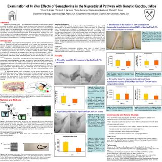
Figure 7: Sections through the dorsomedial hypothalamic nucleus of a Npn 1 loxP/loxP; Th-Cre+ and a Npn 1 loxP/lox P; Th-Cre- brain stained for Th. Note the collection of Th labeled neurons on each side of the ventricle. Npn 1 loxP/loxP; Th-Cre+ Npn 1 loxP/loxP; Th-Cre- Results • A trend for more SNc Th+ neurons in Npn1loxP/loxP; Th-Cre+ brains A Figure 8: Graph comparing the average size of DMN between Npn1loxP/loxP;Th-Cre+ and Npn1loxP/loxP;Th-Cre- brains. Figure 9: Graph showing the average number of Th positive neurons present in the DMN of Th-Cre+ brains compared to Th-Cre- brains. • A trend for more Th+ neurons in the paraventricular hypothalamic nucleus (PVN) of Npn1loxP/loxP; Th-Cre+ brains compared to controls B A Figure 10: Graph comparing the average size of PVN between Npn1loxP/loxP;Th-Cre+ and Npn1loxP/loxP;Th-Cre- brains. Figure 11: Graph showing the average number of Th positive neurons present in the PVN of Th-Cre+ brains compared to Th-Cre- brains. Npn1loxP/wt Npn1loxP/wt X Th-Cre Npn1loxP/loxP X loxP loxP 5’ 3’ Npn1 ZBgeoloxP/wt (GFP) Npn1loxP/loxP Th-Cre X Cre 5’ 3’ loxP loxP 5’ 3’ loxP GFP lacZ Cre Npn1loxP/loxP Th-Cre GFP 5’ 3’ loxP B A Examination of In Vivo Effects of Semaphorins in the Nigrostriatal Pathway with Genetic Knockout Mice 1Chino S. Aneke, 2Elizabeth H. Jackson, 2Tisina Samaroo, 2Claire-Anne Gutekunst, 2Robert E. Gross 1Department of Biology, Spelman College, Atlanta, GA; 2Department of Neurological Surgery, Emory University, Atlanta, GA Abstract Parkinsons Disease (PD) is a neurodegenerative disorder that affects many people worldwide. In patients with PD, there is a loss of dopaminergic neurons in the substantia nigra pars compacta (SNc) located in the ventral midbrain. This loss of dopaminergic neurons leads to a decreased level of dopamine. Our laboratory has preliminary data showing that the semaphorins,a group of axon guidance molecules, are present in the nigrostriatal pathway and therefore participate in its development. However, the exact role of semaphorins in the nigrostriatal pathway has not yet been identified. Here, we use genetic knockout mouse strains to examine the in vivo effects of semaphorins in the development and establishment of the nigrostriatal pathway. Immunocytochemistry: Adult mouse brain tissue sections (N=6 (Npn1loxP/loxP;Th-Cre+) and N=14 (Npn1loxP/loxP;Th-Cre-))were collected and rinsed in PBS three times for 10 minutes each. The tissues were incubated in hydrogen peroxide and 0.1% triton for 20 minutes to rid the cells of peroxidase and permeabilize the tissue, respectively. They were washed in PBS again, in the manner stated above and incubated in a solution of PBS and Normal Goat serum for blocking. The tissues were then washed with PBS, and put in a primary antibody for overnight incubation in a room at 40oC. After, the tissues were rinsed in PBS and incubated in a secondary antibody, a goat anti rabbit IgG, rinsed and incubated in avidin-biotin complex. The tissues were put in a solution of hydrogen peroxide and diaminobenzedene. They were washed in PBS again in the manner stated above and the tissue sections were mounted on gelatin coated slides. Antibodies: Rabbit anti-Tyrosine hydroxylase antibdoes were used to detect Tyrosine hydroxylase-expressing neurons (PelFreeze). Mouse anti-Cre-recombinase antibodies were used to detect Cre-recombinase (Chemicon). • No difference in the number of Th+ neurons in the dorsomedial hypothalamic nucleus (DMN) of Npn1loxP/loxP; Th-Cre+ brains compared to controls Introduction Dopamine, norepinephrine and epinephrine are the main catecholamines that play an important role as neurotransmitters in the central and peripheral nervous systems. Dopamine controls behaviors such as voluntary movement, drug use and abuse, motivation, reward and reinforcement. Tyrosine hydroxylase (TH), the rate limiting enzyme in the synthesis pathway of these catecholamines, converts tyrosine into 3, 4-dihydroxyphenylanine, also known as levodopa (L-DOPA) (Gelman, 2003). L-DOPA is converted to dopamine by an enzyme called L-amino acid decarboxylase (L-AADC). Dopamine is then converted into norepinephrine and then into epinephrine. There are many regions in the brain where catecholaminergic neurons are found (ex: A1 to A17 cell nuclei). Semaphorins are secreted or membrane-bound proteins that play a role in axon guidance in several pathways in the brain. Semaphorins have cell surface receptors that are bound to them with high affinity in these pathways. The known receptor for class 3 semaphorins (sema3A) is neuropilin 1 (npn1) and the co-receptor is plexin A1 (Fujisawa, 1997). Semaphorins bind to npn1 which is bound to its co-receptor, plexin A1. Plexin A1 then provides the signal through the cell membrane to the cell. Although we have localized semaphorins in the nigrostriatal pathway, their role in this pathway has not yet been identified. To determine whether npn1 is necessary for the proper development of the nigrostrital pathway we have crossed several strains of mice to generate a strain lacking the npn1 gene only in the tyrosine hydroxylase expressing cells. We have then compared the development of the nigrostriatal pathway and other dopaminergic pathways between this mice lacking npn1 in TH expressing neurons and normal controls. Figure 4:A) Substantia nigra of a Th-Cre- brain and that of a Th-Cre+ brain immunostained with anti-Th antibodies. B) Graph showing the average number of Th positive neurons present in the SNc of Th-Cre+ brains compared to Th-Cre- brains. There is a trend for more neurons per section in the Th-Cre+ brains. Figure 1:A) Mesolimbic (blue) and nigrostriatal dopaminergic (green) tracts in human brain. B) The development of the nigrostriatal pathway during embryogenesis in the mouse. Legand: Col – Colliculus; SN – Substantia Nigra; Thal – Thalamus; LGE – Persisting Germinal Zone; Str – Straitum. Material and Methods Crosses: Figure 2: A) Schematic showing how the animals were generated. B) Coronal brain sections through the substantia nigra from (Npn1loxP/loxP;Th-Cre-) and (Npn1loxP/loxP;Th-Cre+) adult mice were co-stained for Tyrosine hydroxylaze (TH in green) and Cre-recombinase (in red). Nuclei were stained with Hoescht (blue). TH labeling is found in the cytoplasm of the neurons whereas Cre-recombinase labeling is present in the nuclei as expected. In the (Npn1loxP/loxPxTh-Cre) animals, many of the TH positive neurons are also posititive for Cre-recombinase. Genotyping and tissue preparation: Mice were genotyped by PCR and Cre expression was confirmed by immunocytochemistry. TH Cre TH/Cre/Dapi Npn1loxP/loxP • Significantly wider mfb in Npn1loxP/loxP; Th-Cre+ brains Npn1loxP/loxP Th-Cre • Conclusions and Future Studies: • Our preliminary results suggest that npn1 might participate in the number of Th expressing neurons in SNc and PVN but not in the DMH. • Npn1 also plays a subtle but significant role in restricting the spread of the mfb fibers. • Future studies will include: • ICC for Npn1 to determine the extent of Npn1 depletion caused by Cre expression; • Increase number of animals examined for better statistics; • Examine pathway at earlier developmental stages; • Examine potential behavioral effects of Th specific Npn1 deletion. Figure 5:A) Medial forebrain bundle of an Npn1loxP/loxP;Th-Cre- brain. B) Medial forebrain bundle (mfb) of a Npn1loxP/loxP;Th-Cre+ brain. This bundle is more spread out than that of the Th-Cre- brain. The area of the mfb is outlined for measurement. Figure 6: Graph comparing the average mfb size between Npn1loxP/loxP;Th-Cre+ and Npn1loxP/loxP;Th-Cre- brains. The mfb fiber bundle is significantly larger in the Npn1loxP/loxP;Th-Cre+ compared to Npn1loxP/loxP;Th-Cre- brains. * indicates t-test p=0.019. A B B A Resources This material is based upon work supported by the Howard Hughes Medical Institute under Grant No. 52003727 and by the National Science Foundation Award # 0450303 (Subaward # I-66-606-63 to Emory University). References Gelman et. al. "Transgenic Mice Engineered to Target Cre/LoxP-Mediated DNA Recombination Into Catecholaminergic Neurons". Technology Report 14 May 2007: 196-202. Lindeberg et.al. "Transgenic expression of Cre Recombinase From the Tyrosin Hydroxylase Locus". Technology Report 16 March 2004: 67-73. Fujisawa et.al, "Roles of a neuronal cell-Surfave Molecule, neuropilin, in nerve fiber fasiculation and guidance". Cell & Tissue Research 11 June 1997: 465-470. Novak et. al, "Article". Z/EG, a Double Reporter Mouse LIne That Expresses Enhanced Green Fluorescent Protein Upon Cre-mediated Excision 28 September 2000: 147-155. * Figure 3:A) Genotypes of P1 brains. The results show that the brains are npn-1 homozygous. The brains that have a white band are Th-Cre+ ( 3 & 4), while those that do not (1 and 2) are Th-Cre-. B) Cre staining of a brain section through the SNc of a Th-Cre+ brain. Cre is present in the nuclei of many neurons in the SNc (outlined). Cre-recombinase is responsible for excising the npn1 gene from the Npn1 loxP/loxP strain.


