Download

1 / 27
490 likes | 1.13k Vues
INTESTINAL OBSTRUCTION. Fadi J. Zaben RN MSN. Overview:. The small intestine and colon are components of digestive tract, which processes the foods what we eat.

E N D
INTESTINAL OBSTRUCTION Fadi J. Zaben RN MSN
Overview: The small intestine and colon are components of digestive tract, which processes the foods what we eat. The small intestine and colon extract nutrients from the foods. What isn't absorbed by the small intestine and colon continues along the digestive tract and is expelled as stool during a bowel movement.
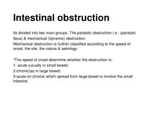
Definition: • Intestinal obstruction occurs when the passage of intestinal contents through the lumen is impaired. • Intestinal obstruction is an interruption in the normal flow of intestinal contents along the intestinal tract. • The block may occur in the small or large intestine, may be complete or incomplete, may be mechanical or paralytic, and may or may not compromise the vascular supply. • Obstruction most frequently occurs in the young and the old.
Bowel obstruction can occur in both the small and large bowel. • The small bowel is most commonly affected, with the ileum as the most common site of obstruction. • Large bowel obstruction accounts for only 15% of cases of bowel obstruction and the sigmoid colon is the most common site of obstruction. • The location of the obstruction, the degree of obstruction, and the presence of ischemia are important distinctions because treatment varies.
Types of Intestinal Obstruction: • Mechanical obstruction. • Paralytic (adynamic, neurogenic) ileus. • Strangulation obstruction.
Mechanical obstruction: • It is a physical block to passage of intestinal contents without disturbing blood supply of bowel. • High small-bowel (jejunal) or low small-bowel (ileal) obstruction occurs four times more frequently than colonic.
Continue…… • Caused by: • Extrinsic adhesions: from surgery, hernia, wound dehiscence, masses, volvulus (twisted loop of intestine). Up to 70% of small bowel obstructions are caused by adhesions. • Intrinsic: hematoma, tumor, intussusception (telescoping of intestinal wall into itself), stricture or stenosis, congenital (atresia, imperforate anus), trauma, inflammatory diseases (Crohn's, diverticulitis, ulcerative colitis)
Continue…… • Intraluminal: foreign body, fecal or barium impaction, polyp, gallstones, meconium in infants • In postoperative patients, approximately 90% of mechanical obstructions are due to adhesions. In nonsurgical patients, hernia (most often inguinal) is the most common cause of mechanical obstruction.
Paralytic (adynamic, neurogenic) ileus: • Peristalsis is ineffective (diminished motor activity perhaps because of toxic or traumatic disturbance of the autonomic nervous system). • There is no physical obstruction and no interrupted blood supply. • Disappears spontaneously after 2 to 3 days.
Continue….. • Causes include: • Spinal cord injuries; vertebral fractures. • Postoperatively after any abdominal surgery. • Peritonitis, pneumonia. • Wound dehiscence (breakdown). • GI tract surgery.
Strangulation Obstruction: • It compromises blood supply, leading to gangrene of the intestinal wall. • Caused by prolonged mechanical obstruction.
Altered Physiology: • Increased peristalsis, distention by fluid and gas, and increased bacterial growth proximal to obstruction. The intestine empties distally. • Increased secretions into the intestine are associated with diminution in the bowel's absorptive capacity. • The accumulation of gases, secretions, and oral intake above the obstruction causes increasing intraluminal pressure. • Venous pressure in the affected area increases, and circulatory stasis and edema result. • Bowel necrosis may occur because of anoxia and compression of the terminal branches of the mesenteric artery. • Bacteria and toxins pass across the intestinal membranes into the abdominal cavity, thereby leading to peritonitis. • Closed-loop obstruction is a condition in which the intestinal segment is occluded at both ends, preventing either the downward passage or the regurgitation of intestinal contents.
Risk Factors: • Diseases and conditions that can increase risk of intestinal obstruction include: • Abdominal or pelvic surgery, which often causes adhesions. • Crohn's disease. • Cancer within your abdomen, especially if their a surgery to remove an abdominal tumor or radiation therapy.
Clinical Manifestations: • Abdominal distention. • Abdominal fullness, gas. • Abdominal pain and cramping. • Breath odor. • Constipation. • Diarrhea. • Vomiting. • Fever, peritoneal irritation, increased WBC count, toxicity, and shock may develop with all types of intestinal obstruction.
Tests and Diagnosis: • Physical exam. • Fecal material aspiration from NG tube • Abdominal and chest X-rays: • May show presence and location of small or large intestinal distention, gas or fluid. • Bird beak lesion in colonic volvulus. • Foreign body visualization.
Continue…… • Contrast Studies: • Barium enema may diagnose colon obstruction or intussusception. • Ileus may be identified by oral barium or Gastrografin. • Laboratory Tests: • May show decreased sodium, potassium, and chloride levels due to vomiting. • Elevated WBC counts due to inflammation; marked increase with necrosis, strangulation, or peritonitis. • Serum amylase may be elevated from irritation of the pancreas by the bowel loop. • Flexible sigmoidoscopy or colonoscopy may identify the source of the obstruction such as tumor or stricture.
Treatment: • Nonsurgical Management. • Surgery.
Nonsurgical Management: • Correction of fluid and electrolyte imbalances with normal saline or Ringer's solution with potassium as required. • NG suction to decompress bowel. • TPN may be necessary to correct protein deficiency from chronic obstruction, paralytic ileus, or infection. • Analgesics and sedatives, avoiding opiates due to GI motility inhibition. • Antibiotics to prevent or treat infection. • Ambulation for patients with paralytic ileus to encourage return of peristalsis.
Surgery: • Consists of relieving obstruction. Options include: • Closed bowel procedures: lysis of adhesions, reduction of volvulus, intussusception, or incarcerated hernia • Enterotomy for removal of foreign bodies. • Resection of bowel for obstructing lesions, or strangulated bowel with end-to-end anastomosis • Intestinal bypass around obstruction • Temporary ostomy may be indicated.
Complications: • Dehydration due to loss of water, sodium, and chloride. • Peritonitis. • Shock due to loss of electrolytes and dehydration. • Death due to shock.
Nursing Assessment: • Assess the nature and location of the patient's pain, the presence or absence of distention, flatus, defecation, emesis, obstipation. • Listen for high-pitched bowel sounds, peristaltic rushes, or absence of bowel sounds. • Assess vital signs.
Nursing Diagnoses: • Acute Pain related to obstruction, distention, and strangulation. • Risk for Deficient Fluid Volume related to impaired fluid intake, vomiting, and diarrhea from intestinal obstruction. • Diarrhea related to obstruction. • Ineffective Breathing Pattern related to abdominal distention, interfering with normal lung expansion. • Risk for Injury related to complications and severity of illness. • Fear related to life-threatening symptoms of intestinal obstruction.
Nursing Interventions: Achieving Pain Relief: • Administer prescribed analgesics. • Provide supportive care during NG intubation to assist with discomfort. • To relieve air-fluid lock syndrome, turn the patient from supine to prone position every 10 minutes until enough flatus is passed to decompress the abdomen. • A rectal tube may be indicated.
Maintaining Electrolyte and Fluid Balance: • Measure and record all intake and output. • Administer I.V. fluids and parenteral nutrition as prescribed. • Monitor electrolytes, urinalysis, hemoglobin, and blood cell counts, and report any abnormalities. • Monitor urine output to assess renal function and to detect urine retention due to bladder compressions by the distended intestine. • Monitor vital signs; a drop in BP may indicate decreased circulatory volume due to blood loss from strangulated hernia.
Maintaining Normal Bowel Elimination: • Collect stool samples to test for occult blood if ordered. • Maintain adequate fluid balance. • Record amount and consistency of stools. • Maintain NG tube as prescribed to decompress bowel.
Maintaining Proper Lung Ventilation: • Keep the patient in Fowler's position to promote ventilation and relieve abdominal distention. • Monitor ABG levels for oxygenation levels if ordered.
Preventing Injury Due to Complications: • Prevent infarction by carefully assessing the patient's status; pain that increases in intensity or becomes localized or continuous may herald strangulation. • Detect early signs of peritonitis to minimize this complication. • Avoid enemas, which may distort an X-ray or make a partial obstruction worse. • Observe for signs of shock. • Watch for signs of (metabolic alkalosis and metabolic acidosis.