Download
1 / 20
201 likes | 545 Vues
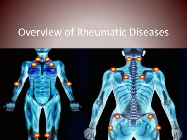
AUTOANTIBODIES IN RHEUMATOLOGY. G. Cooke VTS Trainee. AIMS & OBJECTIVES. Nothing radical Just to enhance consideration of the role of autoantibody titres when… Considering rheumatological diagnosis Assessing for exacerbation / treatment response Interpreting results / letters.

E N D
AUTOANTIBODIES IN RHEUMATOLOGY G. Cooke VTS Trainee
AIMS & OBJECTIVES • Nothing radical • Just to enhance consideration of the role of autoantibody titres when… • Considering rheumatological diagnosis • Assessing for exacerbation / treatment response • Interpreting results / letters
Introduction (1): Autoantibodies?? - Immunoglobulins - Detectable in people with a variety of clinical presentations & in healthy individuals
Introduction (2): - No autoantibody confirms a diagnosis or determines treatment – adjunctive role predominantly - Ask – Will knowing the value of x, y or z alter my management of this patient?
RHEUMATOID FACTOR (1): - IgM which reacts with the fc component of abnormal, antigenic IgG to produce immune complexes complement activation…inflammatory cascade - Present in 1% of healthy individuals - Present in 70-90% of RA patients - Levels increase with age
RHEUMATOID FACTOR (2): - Blood titres >1:80 considered +ve and suggestive of RA - Titres between “unidentifiable” and 1:80 – consider: SLE, Sjogrens (or healthy) - “unidentifiable” titres don’t exclude RA (10-30% RA patients seronegative)
RHEUMATOID FACTOR (3): - High titres severe articular / extra-articular disease in RA - Levels don’t alter significantly with Rx - 80% sensitivity (therefore –ve doesn’t exclude diagnosis) - PPV 50%
RHEUMATOID FACTOR (4): - RF +ve patients? Also consider… - SLE / Sjogrens - Chronic viruses - SBE - TB - Dermatomyositis - Scleroderma - EBV - Leukaemia - Cirrhosis - Syphilis - Renal disease
ANTI-NUCLEAR ANTIBODIES (1): - Strongly associated with SLE (but also other rheumatological conditions, bizarre syndromes and healthy people) - MULTIPLE subtypes of Anti-Nuclear Antibodies (ANA, ds-DNA, ss-DNA, anti-DNP, SS-A, SS-B, Scl-70, RNP, RANA, RAP, Antimicrosomal, Antithyroglobulin, ASMA)
- 95% SLE have +ve ANA (good sens – -ve test good for exclusion of SLE) - Specificity for ANA in SLE low (57% - +ve ANA not great at confirming SLE) [false +ve with MULTIPLE medications] - If lupus personified walks in – check ANA, if a bit grey, probably not worth it - Doesn’t reflect disease activityin SLE (use clinical features / ESR / C3 / C4 / dsDNA) ANTI-NUCLEAR ANTIBODIES (2):
ANTI-NUCLEAR ANTIBODIES (3): - More confusion… Different patterns when studies under UV microscopy and ANA subgrouped accordingly (often referred to in clinic letters / ICE results): Homogenous: SLE / Mixed CTD Peripheral: SLE Speckled: SLE / Sjogrens / Scleroderma Polymyositis / RA / Mixed CTD Nucleolar: Scleroderma / Polymyositis
ANTI-NUCLEAR ANTIBODIES (5): - +ve ANA also a/w… - Chronic Hepatitis - PAN - EBV - Leukaemia - Myaesthenia Gravis - Cirrhosis - Order an ANA when… - Support diagnosis of CTD when suspected clinically (FU with specific tests) - To exclude SLE with 1-2 features and no clear alternative
ANTI-ds-DNA ANTIBODIES: - Less sensitive but more specific for SLE compared to ANA - Very low false +ve rate - Do relate to disease activity – esp. renal involvement (e.g. monitoring lupus nephritis)
ANTI-Sm ANTIBODIES: - AKA Anti-Smith (notsmoothmuscle) - V. SLE specific – but present in 30% cases so not particularly sensitive for SLE - Associated with severity and extent of renal involvement - False +ve a/w EBV – molecular mimicry
ANTI-RNP (ribonuclear protein) ANTIBODIES: - Poor sensitivity and specificity for SLE - Present in 40% SLE patients - High levels diagnostic of Mixed CTD - A/W milder renal disease & Raynaud’s
ANTI-SCLERODERMA ANTIBODIES: - Scl-70 / Scleroderma Abs - Present in 45% with scleroderma (PSS) - Scl-70 titre relates to both likelihood of diagnosis, and disease activity - Absence doesn’t exclude scleroderma - Also a/w: Mixed CTD SLE Sjogren’s RA Polymyositis
Anti-Ro / Anti-La / Anti-SS-C (1): - Confusingly, Anti-Ro = Anti-SS-A Anti La = Anti-SS-B - SS-A / SS-B – used to diagnose Sjogren’s (Prim / Sec) - SS-A in 60-70% Primary Sjogren’s; SS-B in 50% (SS-A + SS-B +ve = confirms diagnosis) - Can differentiate between 1° & 2° Sjogren’s – SS-B found only in 1° disease
Anti-Ro / Anti-La / Anti-SS-C (2): - SS-A found in 25% SLE cases (present in the majority of ANA-ve SLE patients) / SS-B never found in SLE - SS-C +ve in 75% RA patients / RA + 2° Sjogren’s - High Anti-SS levels… Sjogren’s more likely Disease more active - Anti-SS levels fall with treatent
CONCLUSION: - Think before you request - Clinical impression hugely valuable in guiding Ix choice - Sensitive tests first… specific later to confirm diagnoses