Download
1 / 60
610 likes | 1.01k Vues
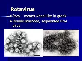
Rotavirus Infection. Children’s Hospital, Zhejiang University School of Medicine Jiang Mizu. What is Rotavirus ? Electron microscopic View of Rotavirus. “ Rota” in Latin means wheel First detected in April, 1973 by R Bishop and team from a biopsy of an

E N D
Rotavirus Infection Children’s Hospital, Zhejiang University School of Medicine Jiang Mizu
What is Rotavirus ?Electron microscopic View of Rotavirus “Rota” in Latin means wheel First detected in April, 1973 by R Bishop and team from a biopsy of an Australian child with severe gastroenteritis. Rotavirus particles in stool filtrate Photo Credit : F.P. Williams, U.S. Environmental Protection Agency; Adapted from Parashar et al, Emerg Inft Dis 1998,14(4) 561–570
What is Gastroenteritis? • Gastroenteritis is second only to respiratory illness as a cause of childhood morbidity worldwide. • Gastroenteritis: nausea, vomiting, diarrhea, abdominal cramping, and fever occur 6-48h after exposure. • Most gastroenteritis is caused by viral infection; bacterial, parasitic, and protozoal illnesses are less frequent but not uncommon.
What is diarrhea? • Definition: increased total daily stool output (> 10g/kg/d), is usually associated with increased stool water content. • Loose consistency(性状改变):watery diarrhea, mucous diarrhea, bloody diarrhea • Increased stool frequency(次数增多) • Duration • Acute (< 14 d) • Persistent (14 d to 2 m) • Chronic (> 2 m)
Rotavirus and diarrhea • Viruses are the most common cause of acute gatroenteritis in developing and developed countries, such as rotavirus, astrovirus, adenovirus, and caliciviruses (Norwalk agent) • Rotavirus, a 67-nm double-stranded RNA virus with at least eight serotypic variants, is the most common. • As with most viral pathogens, rotavirus affects the small intestine, causing voluminous watery diarrhea without leukocytes or blood.
Characteristicsof Rotavirus Appearance of ‘hubbed wheel’ with spokes on electron microscopy 65-75nm diameter Three concentric layers: double capsid (external and internal) and a core (nucleocapsid)
Characteristics of rotavirus Outer Capsid : 2 structural proteins involved in viral adsorption and penetration into epithelial cells. - VP7 : glycoprotein or G protein VP7 VP4 14 G types described Five common VP7 serotypes in humans (G1-G4, G9), but 5 others identified
Characteristics of rotavirus Outer Capsid : 2 structural proteins involved in viral adsorption and penetration into epithelial cells. - VP4 : protease - cleaved or P protein VP7 VP4 20 P genotypes described. One common VP4 genotype P[8] , but four others detected
Characteristics of rotavirus Inner Capsid : VP6 most abundant major group specific antigen Seven groups of Rotavirus (A to G), but only 3 (A,B,C) pathogenic in human Group A strains account for >95% clinical infection. Human group A subdivided into subgroup I and II
Characteristics of rotavirus Core : Viral genome - composed of 11 segments of double- stranded RNA.
A组RV病毒基因组功能 基因片段: 1 2 3 4 6 9 编码 结构蛋白: VP1 VP2 VP3 VP4 VP6 VP7 (核心) (核心) (核心) (外壳) (内壳) (外壳 区分G血清型1-14) 裂解 抗原区分(A-G组) VP5 VP8 A组为Ⅰ,Ⅱ亚群 (P血清型1-44) 基因片段: 5 7 8 10 11 编码 非结构蛋白:NS53 NS34 NS35 NS28 NS26 (NSP1 NSP2 NSP3 NSP4 NSP5)
Developed Countries Less Developed Countries Parasites Unknown Rotavirus Unknown Rotavirus Otherbacteria Escherichia coli Bacteria Adenovirus Adenovirus Astrovirus Calicivirus Astrovirus Calicivirus Epidemiology Epidemiology Distribution of pathogens reported to cause endemic/epidemic gastroenteritis and infantile vomiting and diarrhea From Kapikian AZ, Chanock RM. Rotaviruses. In: Fields Virology 3rd ed. Philadelphia, PA: Lippincott-Raven; 1996:1659.
Introduction Rotavirus • Rotavirus is the most common diarrheal pathogen in children worldwide1 • Globally more than 125 million cases of infantile gastroenteritis2 • 440,000 deaths per year mainly in less developed countries3 Estimated global distribution of 440,000 annual deaths in children <5 years old caused by rotavirus diarrhea31 dot = 1000 deaths 1Parashar et al, Emerg Infect Dis 1998 4(4) 561–570; 2Linhares and Bresee, Pan Am J Public Health 2000 8(5) 305–331; 3Parashar et al, Emerg Infect Dis 2003 9(5) 565–572
Rotavirus-attributable mortality per 1000 children under 5 years of age 0.0-0.1 0.2-0.5 0.6-0.9 1.0-1.9 2.0-3.4
440,000 deaths 2.1 million inpatient visits 25 million outpatient visits 111 million episodes Global rotavirus disease burden Risk Events 5% of all deaths in children < 5 are due to rotavirus 1 : 293 1 : 60 1 : 5 1 : 1 Parashar et. al., Emerg Infect Dis, 2002
Epidemiology • RV infection is most common in winter months in temperate climates. • Disease tends to be most severe in patients 3-24 mo of age, although 25% of the cases of severe disease occur after 2 yr of age, with serologic evidence of infection developing in virtually all children by 4-5 yr of age. • Infants younger than 3 mo of age are relatively protected by transplacental antibody and possibly breast-feeding.
Transmission • Transmission takes place both via the fecal-oral route by contaminated food, water or contaminated toy and from person to person. • Outbreaks are common in children’s hospitals and child-care centers. • Large quantities of virus are shed in the stool during the first week of infection. • The virus survives for hours on hands and for days on environmental fomites.
Pathophysiology of rotavirus infection • In viral infection, diarrhea is noninflammatory and results from an enteropathy in which the death of mature villus-tip cells (responsible for disaccharide digestion and monosaccharide absorption) causes an osmotic diarrhea due to the malabsorption of sugars.
Normal transport of nutrients and electrolytes across the GI tract • The glucose-sodium co-transporter, requires the presence of a sodium gradient across the brush border membrane (Na+- K+ATPase). • The electroneutral NaCl- coupled pathway that involves the double exchange mechanism by the Na+-H+ exchanger (NHE) and the Cl-HCO3- exchanger located at the apical membrane. Jejunum Ileum Colon
Pathogenesis • Viruses that cause human diarrhea selectively infect and destroy villus tip cells in the small intestine. • Biopsies of the small intestine show variable degrees of villus blunting and round cell infiltrate in the lamina propria. • Pathologic changes may not correlate with the severity of clinical symptoms and usually resolve before that clinical resolution of diarrhea.
Pathogenesis • In the small intestine, the upper villus enterocytes are differentiated cells, which have both digestive functions, such as hydrolysis of disaccharides, and absorptive functions, such as the transport of water and electrolytes via glucose and amino acid co-transports. • The crypt enterocytes are undifferentiated cells, which lack the brush border hydrolytic enzymes and are net secretors of water and electrolytes.
Pathogenesis • Selective viral infection of intestinal villus tip cells thus leads to • An imbalance in the ratio of intestinal fluid absorption to secretion • Malabsorption of complex carbohydrates, particularly lactose • Rotavirus nonstructural protein may function as an enterotoxin.
Clinical manifestation of rotavirus infection • The most common cause of acute noninflammatory gastroenteritis in infants and toddlers. • Disease incidence peaks in the cooler fall and winter months (year-round). • The peak age incidence is 3 to 24 months. • The incubation period for RV is 24-48 h. • Vomiting is the first symptom in 80-90% of pts, followed within 24 h by low-grade fever and voluminous watery diarrhea and non bloody.
Clinical manifestation of rotavirus infection • Diarrhea is usually self-limited, abating with 4-8 days but may last longer in young infants or immunocompromised pts. • The white blood cell count is rarely elevated. • The stool does not contain blood or white cells. • Metabolic acidosis results from bicarbonate loss in the stool, ketosis from poor intake, and lactic acidemia from hypotension and hypoperfusion.
Diagnosis • In most cases, a satisfactory diagnosis can be made on the basis of clinical and epidemiologic features. • Enzyme immunoassays, which offer approximately 90% specificity and sensitivity, are available for detection of group A RV in stool samples. • More obscure cases can be studied by electron microscopy of stools, RNA electrophoresis, nucleic acid hybridization, and polymerase chain reaction assays. • Specific identification of rotavirus in not required in every case, especially in outbreaks.
Stools studies Findings Implications Gross examination Blood, mucus, pus Bacterial infection Microscopic examination >5 WBC/hpf Bacterial infection Chemical examination • Stool pH pH<5 Viral infection, Carbohydrate malabsorption • Stool-reducing substances + Viral infection, Carbohydreate malabsorption
Determination of hydration status • The most common causes of dehydration in children are vomiting and diarrhea. • Dehydration is classified by the percentage of total body water lost: mild (<5%), moderate (5-10%), and severe (>10%). • A variety of signs and symptoms and ancillary date help to estimate the degree of dehydration.
Degree of dehydration Clinical signs mild moderate severe Decrease in body weight 3-5% 5-10% 10-15% Skin Turgor normal decreased Markedly decreased Color normal pale markedly decreased Mucous membranes Dry Mottled or gray; parched Hemodynamic signs Pulse normal slight increase tachycardia Capillary refill 2-3 s 3-4 s >4 s blood pressure normal low perfusion normal circulatory collapse Fluid loss urinary output mild oliguria oliguria anuria Tears Decreased absent Urinary indices specific gravity >1.020 anuria Urine [Na+] <20mEq/L anuria
Normal values of an arterial blood gas pH 7.35-7.45 PO2 80-100mmHg PCO2 35-45mmHg [HCO3-] 20-28mmol/L SBE -3-3mmol/L Na+ 135-145mmol/L K+ 3.5-5.5mmol/L Cl- 96-108mmol/L Mg + 0.62-0.94mmol/L
Electrolyte Disorders • Sodium disorders Isotonic dehydration: [Na+] 130-150mmol/L Hypotonic dehydration: [Na+] <130mmol/L Hypertonic dehydration: [Na+] >150mmol/L • Potassium disorders Hyperkalemia: [K+] >5.5mmol/L Hypokalemia: [K+] <3.5mmol/L
Metabolic Acidosis According to AG= [Na+] - ([HCO3-] + [Cl-]) • Normal type: 8-16mmol/L [HCO3- ] • Increased type: >16mmol/L [H+] According to [HCO3-] • Mild [HCO3-] 18-13mmol/L • Moderate [HCO3-] 13-9mmol/L • Severe [HCO3-] <9mmol/L
Differential diagnosis • In infancy, the differential diagnosis of acute gastroenteritis includes diarrhea associated with other infections such as urinary tract infection, otitis media, sepsis, and pneumonia. • Depending on the geographic location, enteric adenoviruses or caliciviruses are the next most common viral pathogens in infants. • Other potentially pathogenic viruses include astroviruses, corona-like viruses, Coxsackis viruses, and other small round viruses.
Norwalk virus • The Norwalk agent, a calicivirus, is a small RNA virus that causes epidemic outbreaks of gastroenteritis • Norwalk virus affects school-age children, adolescents, and adults. • After a 24-h incubation period (range,12-72h), patients characterized by fever, vomiting, diarrhea, and often malaise and myalgias. • Stools are loose, watery, and without blood, mucus, or leukocytes. • The duration of symptoms is short, usually 12-60 hours.
Management • The goals • Recognition, prevention, and treatment of dehydration • Maintenance of the nutritional status of the patients. • Supportive treatment • Replacement of fluid and electrolyte deficits and ongoing losses is critical, especially in small infants. • The use of oral rehydration fluid is appropriate in most cases.
Management • No role for antiviral drug treatment. • No benefit for antibiotics • No benefit from antiemetics or antidiarrheal drugs, and there is a significant risk of serious side effects. • Antimotility agents should be avoided • Probiotic organisms such as lactobacillus species has been shown to reduce somewhat the intensity and duration of illness.
Oral rehydration solution (ORS) 配方 成分 (低渗) g/L (标准) g/L 氯化钠 2.6 3.5 无水葡萄糖 13.5 20 氯化钾 1.5 1.5 枸椽酸钠 2.9 2.9 总重量 20.5 27.9
低渗ORS的常用配方 *由于含有更多的枸橼酸盐,口味相对更酸甜,容易被儿童接受
Management of dehyration • The management priorities are stabilization of the patient’s vital signs, replenishment of the intravascular volume, and correction of electrolyte abnormalities • A patient with mild or moderate dehydration can be orally rehydrated if willing and able to tolerate fluids. • IV fluid restoration is necessary for severe dehydration, shock, or if the patient is unable to take fluids orally or has and altered metal status.
Mild and moderate dehydration • Start oral rehydration with an electrolyte solution (ORS), giving a total volume of 30-50 ml/kg over a 3-4 h period. • Failure of oral rehydration is an indication for IV rehydration. • Once the initial rehydration is tolerated, resume giving milk to an infant, whether breast- or formula-fed. • An infant who has large, watery stools can have the milk feedings supplemented with feedings of oral electrolyte solution.
Severe dehydration • Initial intravascular restoration: Give fluid resuscitation with a 20-ml/kg bolus of normal saline (NS) over 20 to 30 min. • Assessing their fluid status (renal or cardiac disease, sickle cell disease) • Obtain blood for electrolytes, blood urea nitrogen (BUN), creatinine, glucose, and urinalysis • Reevaluate the patient after the first bolus, if there is a poor response to the initial bolus, repeat the infusion. • If there is a poor response to two IV boluses, consider other associated organ disease or the need for central venous monitoring before giving a third bolus.
定输液种类 • 等渗脱水:补1/2张液 (2:3:1液或1:1液) • 低渗脱水:补2/3张液 (4:3:2液) • 高渗脱水:补1/5~1/3张液 (1:4液或1:2液) • 临床上判断脱水性质有困难时,可按等渗脱水补给。
Metabolic acidosis • Intravenous NaHCO3 administration may be considered in the setting of moderate-severe metabolic acidosis. • The dose (in mEq) of 5% NaHCO3 : Weight (kg) × Base deficit × 0.3 • The dose (in ml) of 5% NaHCO3: Weight (kg) × Base deficit × 0.5 • Given an half of calculation as a continuous infusion over 1 h. • The effect of NaHCO3 in lowering serum potassium and ionized calcium concentrations must also be considered.
常用的溶液 一、非电解质溶液(无张力溶液) • 5%Glucose • 10%Glucose 二、电解质溶液 • 10%NaCl 11个张力0.9%(等张) • 5%NaHCO3 3.5个张力1.4%(等张) • 11.2%乳酸钠6个张力1.87%(等张)