
Evaluation of Midazolam Pharmacokinetics in Pregnancy Utilizing a Semi-Mechanistic Model
This study presents a semi-mechanistic pharmacokinetic model for midazolam in pregnancy, incorporating hepatic and intestinal metabolism. The model is validated using clinical data and sensitivity analyses to evaluate fetal metabolism, hepatic blood flow, and CYP3A activity. By adapting the model to physiological changes during pregnancy, it provides insights into midazolam disposition.

Evaluation of Midazolam Pharmacokinetics in Pregnancy Utilizing a Semi-Mechanistic Model
E N D
Presentation Transcript
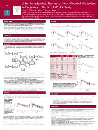
A Semi-mechanistic Pharmacokinetic Model of Midazolam in Pregnancy: Effects of CYP3A Activity Sara K. Quinney1, Mary F. Hebert2, Lang Li3 1 Dept. of OB/GYN, Division of Clinical Pharmacology, Indiana University School of Medicine, Indianapolis, IN; 2University of Washington School of Pharmacy, Seattle, WA; 3Dept. of Medical and Molecular Genetics, Center for Computational Biology and Bioinformatics, Indiana Institute of Personalized Medicine Indiana University School of Medicine, Indianapolis, IN Background Results Historically, physiologically based pharmacokinetic (PBPK) models of pregnancy were used by toxicologists to estimate the concentration of toxicants, including drugs, in various tissues. However, these complex models are difficult to validate with data from clinical trials and pose identifiability issues. We have proposed a simpler, semi-mechanistic PK model, based on the classical two compartment PK model, which incorporates hepatic and intestinal drug metabolism. This approach allows the use of readily-available compartmental PK parameters from the literature or fit from clinical study data. Metabolic enzyme activity is estimated from in vitro rate constants, and optimized to fit clinical data. This model has been validated for midazolam using data from drug-drug interaction studies with ketoconazole, clarithromycin, diltiazem and erythromycin in healthy volunteers [2-4]. Figure 3.Sensitivity Analysis was performed by varying individual parameters 2-fold up (blue line) or down (red line) from initial model (black line). Fetal Vmax was increased 20-fold (blue line) and reduced to 0 (red line). Filled circles represent mean maternal plasma concentrations from Hebert et al. [1]. Objectives This semi-mechanistic PK model is adapted to describe the disposition of midazolam during pregnancy using literature data on the physiological changes that occur during pregnancy. Clinical data obtained following dosing with midazolam [1] is used to further calibrate the model. Sensitivity analyses are performed to illustrate the impact of fetal metabolism, changes in hepatic blood flow, and CYP3A activity. Methods The semi-mechanistic PK model for midazolam in non-pregnant healthy subjects has been reported previously [2-4]. For the pregnancy model, two additional compartments were added to represent the placenta and fetus (Figure 1). Figure 1. Semi-mechanistic PK model for midazolam in pregnancy. Table 1. Effect of varying parameters on AUC, Cmax, and model fit (residual sum of squares) Figure 4. Effect of fetal metabolism on fetal plasma concentrations. Fetal Vmax was increased 20-fold (blue line) or set equal to 0 (red line). Initial model parameters were based midazolam parameters from literature and healthy volunteer data, with alterations in volume of distribution and clearance parameters reflecting the physiological changes associated with pregnancy. In addition to maternal metabolism, midazolam clearance occurs through the fetal liver. Fetal liver metabolism was assumed to be entirely due to CYP3A7 and was estimated by scaling up in vitro Vmax and Km values to estimate Vmax in the fetal liver using the equation [5]: Vmax,Fet =Vmax,3A7 x ISEF x CYP3A7 abundance x MPPGL x Liver Weight Where Vmax,3A7 is the maximal velocity determined in rCYP3A7 in vitro (4 nmol/min/nmol [6]), ISEF is an inter-system extrapolation factor [7] and was set to 1. CYP3A7 abundance is the amount of CYP3A7 enzyme per mg microsomal protein (0.3 nmol per mg [8]), and MPPGL is the mg of microsomal protein per gram of liver (assumed to be equivalent to adult liver, 32 mg/g [9]). Fetal liver weighs 130 g at 38 weeks gestation [10]. Thus, Vmax,Fet is 108 mg/h. The Km for 1’- hydroxymidazolam formation by CYP3A7 is 114 µM [6]. Differential equations were solved using the ODE package in R (v 2.9.2). Model parameters were then calibrated based on mean plasma concentration-time curves obtained from Hebert et al. [1]. Optimum model parameters were identified based on minimization of the residual sum of squares. Subsequent sensitivity analyses were carried out to identify the impact of varying parameters on maternal plasma concentration. Figure 5.Final model of midazolam in pregnancy. Parameters were calibrated based on mean data from Hebert et al. [1]. Black line indicates mean of 500 simulated individuals, grey lines are 5th and 95th confidence intervals. Black circles indicate individual observed maternal plasma concentrations and blue circles indicate mean observed maternal plasma concentrations. Results Discussion The original midazolam model, developed in healthy volunteers, was adequately able to predict midazolam plasma concentrations following a 2 mg oral dose in post-partum women (Figure 2A). Application of the initial model of pregnancy indicates that the bioavailability of midazolam was under-predicted (Figure 2B) • This semi-mechanistic pharmacokinetic model closely depicts the disposition of oral midazolam in women 26-30 weeks of gestation. • Optimization of the model parameters indicates that hepatic, but not intestinal, CYP3A activity is increased during pregnancy. Additional studies are needed to verify this finding. • Sensitivity analysis indicates that fetal metabolism has little impact on the maternal plasma concentration of midazolam. Thus, future models to predict maternal midazolam plasma concentrations in pregnancy may not require inclusion of fetal metabolism. Figure 2. Predicted and observed midazolam concentrations postpartum (A) and during pregnancy (B). Simulated plasma-concentration time profiles in 500 individuals are indicated by the grey lines. Observed maternal plasma concentrations are indicated by triangles (postpartum) and circles (pregnant). References 1. Hebert, M.F., et al., Effects of pregnancy on CYP3A and P-glycoprotein activities as measured by disposition of midazolam and digoxin: a University of Washington specialized center of research study. Clin Pharmacol Ther, 2008. 84(2): p. 248-53. 2. Quinney, S.K., et al., Physiologically Based Pharmacokinetic Model of Mechanism-Based Inhibition of CYP3A by Clarithromycin. Drug Metabolism and Disposition, 2010. 38(2): p. 241-248. 3. Chien, J.Y., et al., Stochastic prediction of CYP3A-mediated inhibition of midazolam clearance by ketoconazole. Drug Metab Dispos, 2006. 34(7): p. 1208-19. 4. Zhang, X., et al., Semiphysiologically based pharmacokinetic models for the inhibition of midazolam clearance by diltiazem and its major metabolite. Drug Metab Dispos, 2009. 37(8): p. 1587-97. 5. Howgate, E.M., et al., Prediction of in vivo drug clearance from in vitro data. I: Impact of inter-individual variability. Xenobiotica, 2006. 36(6): p. 473-497. 6. Williams, J.A., et al., Comparative metabolic capabilities of CYP3A4, CYP3A5, and CYP3A7. Drug Metab Dispos, 2002. 30(8): p. 883-891. 7. Proctor, N.J., G.T. Tucker, and A. Rostami-Hodjegan, Predicting drug clearance from recombinantly expressed CYPs: intersystem extrapolation factors. Xenobiotica, 2004. 34(2): p. 151-78. 8. Shimada, T., et al., Characterization of microsomal cytochrome P450 enzymes involved in the oxidation of xenobiotic chemicals in human fetal liver and adult lungs. Drug Metab Dispos, 1996. 24(5): p. 515-22. 9. Barter, Z.E., et al., Scaling factors for the extrapolation of in vivo metabolic drug clearance from in vitro data: reaching a consensus on values of human microsomal protein and hepatocellularity per gram of liver. Curr Drug Metab, 2007. 8(1): p. 33-45. 10. Basic anatomical and physiological data for use in radiological protection: reference values. A report of age- and gender-related differences in the anatomical and physiological characteristics of reference individuals. ICRP Publication 89. Ann ICRP, 2002. 32(3-4): p. 5-265. Table 1 and Figure 3 show the impact of various parameters on model fit. Vmax,H, Vmax,GW, fu, and ka had large effects on the shape and fit of the curve. However, exclusion of the fetal compartment or increasing the rate of midazolam clearance by the fetus, had little effect on the disposition of nifedipine in maternal plasma or concentrations in fetal plasma (Figure 4). Model calibration indicates that midazolam hepatic Vmax increased 2-fold, as anticipated. However, clearance in the gut wall was similar to the non-pregnant state. In addition, the Ka was increased in the pregnancy model as compared to the healthy volunteer parameters (2.5 vs 1.17h-1). Final model predictions are shown in Figure 5.


