Download
1 / 27
280 likes | 316 Vues
Learn about the principles and techniques of protein immuno-blotting, including SDS-PAGE, Western blotting, and antibody structure. Discover how to visualize and analyze specific proteins using different detection methods. References included.

E N D
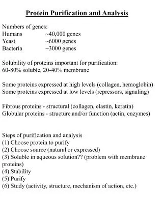
Assiut University Protein immuno-blotting, detection and analysis By prof. Abo Bakr Eltayeb
Electrophoresis • Cation = positively charged ion, it moves toward the cathode (-) • Anion = negatively charged ion, it moves toward the anode (+) • Amphoteric substance = can have a positive/negative/zero charge, it depends on conditions • Principle: • Some substances have different net charges and can be separated into several fractions in external electric field. • But velocity of a particle also depends on the: • size, shape of the particle and given applied voltage
Stacking gel Resolving or separating gel The actual bands are equal in size, but the proteins within each band are of different sizes.
SDS-PAGE,sodium dodecyl sulfate polyacrylamide gel electrophoresis (Laemmli 1970) SDS (sodium dodecyl sulfate) is a detergent (soap) that can dissolve hydrophobic molecules but also has a negative charge • Therefore, if a cell is incubated with SDS, the membranes will be dissolved, all the proteins will be solubalized by the detergent and all the proteins will be covered with many negative charges.
Protein gel (SDS-PAGE) that has been stained with Coomassie Blue.
Western blot It is a jock????? Sir prof. Edwin Southern Inventor of southern blot
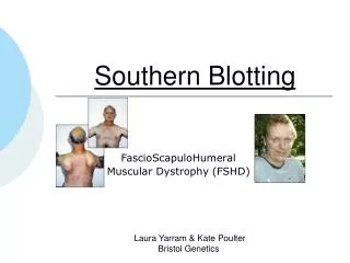
Terminologies.. The Western blot(alternatively, proteinimmunoblot) is an analytical technique used to detect specific proteins in a given sample of tissue homogenate or extract. A Southern blotis a method routinely used in molecular biology for detection of a specific DNA sequence in DNA samples. The northern blotis a technique used in molecular biology research to study gene expression by detection of RNA. Southwestern blotting, based along the lines of Southern blotting (which was created by Edwin Southern) and first described by B. Bowen and colleagues in 1980, is a lab technique which involves identifying and characterizing DNA-binding proteins (proteins that bind to DNA).
Western Blotting (WB) WB is a protein detection technique that combines the separation power of SDS PAGE together with high recognition specificity of antibodies An antibody against the target protein could be purified from serum of animals (mice, rabbits, goats) immunized with this protein Alternatively, if protein contains a commonly used tag or epitope, an antibody against the tag/epitope could be purchase from a commercial source (e.g. anti-6 His antibody)
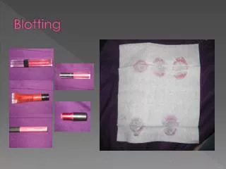
WB: 4 steps • Separation of proteins using SDS PAGE • 2. Transfer of the proteins onto e.g. a nitrocellulose membrane (blotting) • 3. Immune reactions • 4. Visualization
Transfer Wet Semi-dry
Types of membranes Nitrocellulose (NC) high binding capacity, works well with both protein and DNA not need methanol to preparation. Polyvinylidene difluoride (PVDF) high capacity and stable, need methanol for preparation. These both membranes bind proteins non-covalently.
Blocking 5% non-fat milk or BSA with Tween 20: Prevents the primary antibody from binding randomly to the membrane After blocking apply your first Ab at the specific concentration, learn how…? Wash carefully, apply secondary Ab HRB conjugated, wash carefully, detect your specific protein by detection reagent.
Antibody Structure Ig domain: 110 amino acids; globular domain used in many proteins. Variable domains, Constant domains,Hinge. Fab: fragment antigen binding Fc: fragment crystallizable (effector functions)
Immunoglobulins (Ig) are glycoproteins made up of light (L)and heavy(H) polypeptide chains. The simplest antibody molecule has a Y shape and consists of four polypeptide chains:two H chains and two L chains. The four chains are linked by disulfide bonds.
Method for visualization of western blot Most famous 1- colorimetric, by substrate ( ex. DAB) that affected by atomic O resulted from H2O2 hydrolysis by HRB enzyme linked to secondary antibody and give colour 2- ECL, a reagents that affected by atomic O and give luminescence that filmed on X ray films in a dark room, more sensitive that colorimetric method
Comparison between ECL and DAB detection methods DAB ECL ECL is more sensitive 3-5 folds
Look carefully , is your protein found in coomassie blue stain? SDS page stained with coomassie blue The same but after Western blot
Western blot application HIV test Proteins are transferred (blotted) onto the surface of a membrane HIV lysate proteins are separated by size using gel electrophoresis Strips are incubated with patient serum and antihuman IgG conjugated with an enzyme (and chromagen) The membrane is cut into strips
HIV Western Blot Banding Pattern env gp160 gp120 gp 41 gag p55 p18 p24 pol p65 p51 p31
Detection of enzyme activity by the colored substrate in SDS page
References Introduction to Biotechnology by W.J. Thieman and M.A. Palladino. Pearson & Benjamin Cummings 2nd edition. http://www.toodoc.com/SDS-PAGE-ppt.html http://www.bio.davidson.edu/courses/genomics/method/Westernblot.html