Cardiovascular Practical
Explore and identify various cardiovascular structures on microscope slides, including ECG recordings to analyze heart conditions like tachycardia, bradycardia, and fibrillation. Discover the anatomy of vital organs such as the heart, endocrine glands, and hepatic portal system. Study blood vessel types, lymphatic system, and the lower respiratory tract. Ideal for educational purposes and medical training.

Cardiovascular Practical
E N D
Presentation Transcript
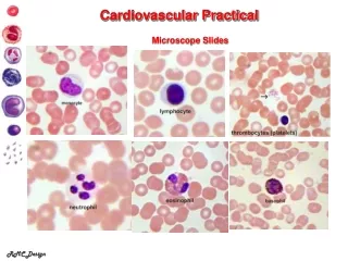
Cardiovascular Practical Microscope Slides RMC Design
ECG Recording What is occurring at A, B, C? Normal Sinus Tachycardia Bradycardia Ventricular fibrillation Ventricular tachycardia Asystole RMC Design
Sheep’s Heart Tricuspid valve RMC Design
Endocrine Glands Pineal gland Hypothalamus Pituitary Gland Thyroid gland Parathyroid glands Thymus Adrenal glands Pancreas Islets of Langerhans Ovaries Testes
Pulse Points Make sure you also Indicate left and right with the arteries RMC Design
HepaticPortalSystem gastric vein splenic vein hepatic portal vein inferior mesenteric vein superior mesenteric vein RMC Design
Circle of Willis labeling anterior communicating a r. middle cerebral a. l. anterior cerebral a. l. posterior communicating a. r. internal carotid artery l. posterior cerebral a. basilar artery a. r. vertebral artery a. RMC Design
Coronary CirculationVessels Coronary Veins great cardiac vein coronary sinsus middle cardiac vein small cardiac vein left coronary artery circumflex a. right coronary a. anterior interventricular a.(LAD) posterior interventricular a. Coronary Arteries marginal a. RMC Design
Blood Vessel Types Artery lumen tunica media (smooth muscle) tunica externa (adventitia)/fibrous CT tunica interna (intima)/endothelium endothelium (simple squamous) Capillary Vein RMC Design
Lymphatic System tonsils l. cervical lymph nodes right lymphatic duct thoracic duct l. subclavian vein thymus r. axillary lymph nodes cisterna chyli spleen r. supratrochlear lymph nodes Peyer’s patches l. inguinal lymph nodes bone marrow lymph vessels
Lymph Node trabecula germinal centers capsule subscapsular sinus afferent lymph vessels afferent lymph vessels cortex hilum follicle medulla sinus efferent lymph vessels
Lower Respiratory Tract thyroid cartilage cricoid cartilage Trachea clavicle r. superior lobe r. primary bronchus Carina (tracheal bifurcation) l. primary bronchus mediastinum l. secondary (lobar) bronchi r. parietal pleura l. tertiary (segmental) bronchus r. visceral pleura r. middle lobe r. inferior lobe r. pleural cavity diaphragm
Vocal Cords base of tongue epiglottis false vocal cords true vocal cords glottis corniculate cartilage