Download
1 / 1
20 likes | 157 Vues
Isolation, Detection + Cellular and Molecular Characterization of Circulating Tumor Cells in Breast Cancer Patients. Introduction:

E N D
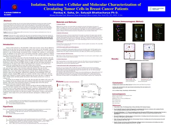
Isolation, Detection + Cellular and Molecular Characterization of Circulating Tumor Cells in Breast Cancer Patients Introduction: Cancer is often referred to as “the silent killer.” At the onset of cancer, cancer cells are difficult to identify; by the time they have been detected, not much can be done to cure the patient. Cancer is characterized as abnormal cells that divide and multiply at an uncontrollable rate. It can be genetically inherited or may develop through environmental factors. Examples that contribute to environmental factors are inhalation of toxic fumes such as insecticides & pesticides, extensive exposure to radiation (UV Rays from the sun and other sources), the over intake of foods with high lipid content, and the construction of nuclear power plants. Tumors may develop in any organ of the body. Eventually the cancer cells metastasize and evade other organs of the body. In general, cancer cells can travel in the body through two ways, the lymphatic system and the bloodstream. The lymphatic system is the body’s secondary immune system and the bloodstream is where red blood cells (RBC) circulate to supply the body with oxygen and nutrients. As cancer progresses the tumor is more visible to the patient because it increases in size. By the time a tumor forms into a small lump, there are more than a billion cancer cells there. Treatments should be taken by the patient to prevent the progression of the tumor. Individuals with cancer may have surgery, where the tumor is physically removed. They may also take chemotherapy, where anticancer drugs are used for treatment. Biological therapy can be used to stimulate cells to restore their ability to fight infections and other diseases. Radiation therapy is an alternative option; It uses high-energy radiation from computerized axial tomography (CAT scan), magnetic resonance imaging (MRI), gamma rays, neutrons, and other sources to kill cancer cells and shrink tumors. Exposure to extensive amounts radiation may also causes side affects. It suppresses the body’s immune system and makes patient more prone to other diseases; patients may even die of common cold. Treatments do not eliminate all of the cancer cells; they just reduce the number of the cancer cells. The remaining cells become dormant and go into circulation in the blood; they carry out normal cell functions. These cancer cells in the blood are known as circulating tumor cells (CTC) and are characterized by the type of molecular marker they have. Tumor markers are branches in specific cells that can often be detected in higher-than-normal amounts in the blood of patients with a certain type of cancer. There is a specific molecular marker of every type of cell; it is these markers that help differentiate, characterize, and distinguish one cell from another. The molecular markers for breast cells are Mammoglobin and cytokeratin-19. Tumor markers are produced by the tumor itself or by the body in response to the presence of cancer or non-cancerous conditions. Tumor marker levels can be useful in the detection and diagnosis of different types of cancer. Pankaj K. Saha, Dr. Satyajit Bhattacharya Ph D. Minimal Residual Lab, Memorial Sloan-Kettering Cancer Center, New York City, NY, 10021, U.S.A Acknowledgments • Memorial Sloan-Kettering Cancer Research Center • Harlem Children Society • Manhattan Center for Science and Mathematics • Dr. Sat Bhattacharya • Mrs. Charlene Chan • Mr. Carlos Franco Harlem Children Society • Materials and Methods: • 1. Specimen collection: • Highly progressive metastatic breast cancer patients were analyzed for the study. All the patients were positive for metastases by imaging, bone scan, and CT scan. A group of healthy individuals with no apparent malignancies were taken as negative controls. Informed consent from all the patients prior to participation in the study was obtained. The peripheral blood was mixed with appropriate amount of phosphate buffered saline (PBS), and separated using Ficoll gradient by density gradient centrifugation. The mononuclear and a portion of the granulocytic layer were taken for further study. • 2. Epithelial Cell Enrichment: • One half of the mononuclear cell fraction isolated from the blood samples was incubated with magnetic beads coated with Ber-EP4 antibody directed against human epithelial antigen, a membrane antigen widely expressed in epithelial cells. The other half of the mononuclear cell fraction (used for microscopic evaluation) was incubated with magnetic beads coated with Ber-EP4 antibody linked to the magnetic beads via a DNA linker. The magnetic cell fraction was isolated and treated with DNase to remove the magnetic beads from the cells. The isolated Immunomagnetic bead-free cells were then cytospun, fixed in Methanol: Acetone (1:1), and stored at -80°C. • 3. RNA Isolation: • Messenger RNA was isolated using Oligo (dT) attached to magnetic beads after epithelial cell enrichment. The isolated RNA was stored at -80°C until further used. • 4. RT-PCR for Specific mRNAs and Gel Electrophoreses: • Single tube, nested RT-PCR for Mammoglobin mRNA was performed on the RNA samples. The PCR amplified products were run on a 2% ethidium bromide agarose gels and observed under a UV transilluminator. To confirm the presence of amplifiable RNA, every sample was subjected to RT-PCR for actin mRNA, and confirmed on agarose gel electrophoresis. • 5. Positive and Negative RT-PCR Controls: • MCF-7 (Breast cell line) and Carcinoma tissue from Breast Cancer Patients were used as positive controls. Negative control PCR consisted of reactions mixtures without any template. • 6. Sensitivity Experiments: • MCF-7 (breast cell line) was used to define the sensitivity of the assays. The cell line was grown and maintained in tissue culture. The MCF-7 cells were mixed with whole blood samples from healthy volunteers to generate serial dilutions. Subsequently, they were processed for further RNA analysis. • 7. Immunohistochemistry (IHC) and Immunofluorescence (IMF): • The slides were processed either by IHC of by IMF. Slides processed for IHC were treated with mouse anti human Mammoglobin, antibody (Hybritech) after pretreatment with Citric acid solution. They were then stained with DAB after treatment with horse anti mouse, IgG and peroxide conjugated with streptavidin. These cells were observed under light microscopy after counter staining with Hematoxilin. • The other group of slides processed for cell cycle evaluation were treated with Hybritech anti – Mammoglobin antibody and then with rabbit anti mouse F(ab’)2-FITC. These were then treated with Propidium Iodide (5 ug/ml) in the presence of RNAseA (100 ug/ml) for 30 minutes at room temperature in the dark. The fluorescence – green (tyrosinase) and red (PI, DNAcontent) was analyzed under a Laser Scanning Microscope Cytometer (LSMC) (Compucyte Corp., Cambridge, MA). Abstract: It is my hypothesis that circulating breast cells can be detected, isolated and characterized from breast cancer patients. Six highly progressive metastatic breast cancer patients were analyzed for this study. The breast cells were isolated by a Novel sensitive Immunomagnetic Method from the mononuclear fraction isolated from leukapheresis. Nested RT-PCR for Mammoglobin was performed. Breast cells were then used for microscopic evaluation, and were enriched immuno-magnetically and cytospun. Slides processed by immunoperoxidase (IPX) were evaluated for the detection of Mammoglobin. Other slides were stained with propidium Iodide (PI, DNA Content red) and subjected to immunofluorescence for Mammoglobin (green) and analyzed by a laser Scanning Microscope Cytometer. Results: Detectable levels of Mammoglobin mRNA was observed in 3 out of every 6 patients, and detectable levels of Actin was observed in 6 out of every 6 patients. Conclusion: We have been able to isolate circulating breast cells using a novel immunobead technique for microscopic evaluation by IPX, LSMC, and RT-PCR. Use of multiple markers greatly enhances the Molecular detection of circulating breast cells. Mammoglobin seems to play a critical role in the proliferation in breast cancer. This technique can be used for Molecular Characterization of the CTC cells in Breast and other solid tumors. This would help Assess, Stratify, and Design Targeted Therapy for Cancer Patients. Pictures (Immunomagneic Method): Results: Immunobead RT-PCR in Breast Cancer Patients Actin PCR B/BM: 154bp Mammoglobin PCR: 215bp Of the six patients that were Out of the 6 tested only 3 of tested, all six had Actin them had Mammoglobin Pictures (Guanidium Isothicyanide Method): Conclusion: (i) Magnetic phase method is more efficient than the Guanidium Isothicyanide Method Using Ultraspec (ii) Cancer cells can be detected at early stages (treatment and monitoring of the cancer patients can be enhanced and customized) (iii) Less radiation therapy, chemotherapy, and immunotherapy • Objectives • (i.) To isolate Circulating breast cells from Peripheral blood of carcinoma patients with breast cancer using a Novel Immunobead Technique for microscopic evaluation by Immunohistochemistry and for Laser Scanning Microscope Cytometer by Immunofluorescence. • (ii.) To detect mRNA by Immunobead RT-PCR. • Hypothesis • Positive selection of epithelial cells using immunobead enrichment of peripheral blood samples from breast cancer patients allows: • (i.) Detection and estimation of breast cancer cells using RT-PCR for Mammoglobin mRNA by Immunohistochemistry (for Light Microscopic examination), and by Immunofluorescence (for Laser Scanning Microscopic Cytometer). • (ii.) Detection of Mammoglobin by RT-PCR • Principles • (i) Positive Selection of epithelial cells is possible using Immunomagnetic beads coated with Ber-EP4 monoclonal antibody directed against the human epithelial antigen, expressed on most normal and neoplastic human epithelial tissues. • (ii) Messenger ribonucleic (mRNA) is very unstable in extracellular environment; therefore, detection or mRNA should indicate the presence of tumor cells in the examined tissue of body fluid. • (iii) PolyA + mRNA can be isolated from selected cells using Immunomagnetic beads coated with Oligo (dT) • References • Cotran, Kumar, Collins: The Pathological Basis of Disease 6th Edition (W.B. Saunders Company) • Foss AJ, Guille MJ, Occleston NL, Hykin PG, Hungerford JL, and LightmanS: The detection of melanoma cells in peripheral blood by reverse transcriptase polymerase chain reaction. Br J Cancer 72: 155 – 159, 1995 • Ghossein RA, Bhattacharya Coit DG. Reverse transcriptase polymerase chain reaction (RT-PCR) detection of melanoma-related transcripts in the peripheral blood and bone marrow of patients with malignant melanoma. What have we learned? Recent Results Cancer Res. 2001;158:63-77. • Ghossein RA, Bhattacharya S.: Molecular detection and characterization of circulating tumour cells and micrometastases in solid tumours. Eur J Cancer. 2000 Aug 36 (13 Spec No):1681-94. • Pelkey TJ, Frierson HF and Bruns DE: Molecular and immunological detection of circulating tumor cells and micrometastases from solid tumors. Clin Chem 42:1369 – 1381, 1996 • Ronald A. Ghossien, Leo Carusone, and Satyajit Bhattacharya: Molecular detection of micrometastases and circulating tumor cells in melanoma prostatic and breast carcinomas. In vivo 14: 237 – 250, 2000