
mlg
E N D
Presentation Transcript
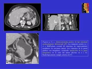
a b Figuras a, b y c. Adenocarcinoma gástrico de tipo intestinal moderadamente diferenciado pT2. Las imágenes axiales (a y c) y RMP-plano coronal (b) muestran un engrosamiento neoplásico en curvatura menor. Los contornos de la pared gástrica son irregulares, existiendo rarefacción de la grasa perigástrica en la zona del tumor (flechas en a y b). Radiológicamente cumple criterios de T3. c
§ * a b Estas figuras muestran la correlación radio-patológica del tumor: las glándulas neoplásicas (cabezas de flecha en b) atraviesan la capa muscular (§) pero sin atravesar la serosa (flecha negra). Interpuestas se identifican fibras de colágeno por la reacción desmoplásica (*) e infiltrado inflamatorio. La zona de rarefacción de la grasa (flecha en a) correspondía a tejido adiposo con áreas de hemorragia e inflamación (flechas negras en c). c