Download
1 / 16
160 likes | 425 Vues
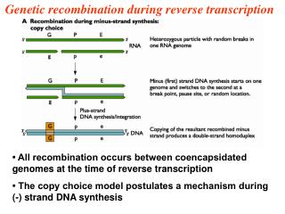
Explore the intricate mechanisms of genetic exchange in bacteria through transformation, transduction, and conjugation. Discover how bacterial recombination leads to adaptability and the generation of diverse strains.

E N D
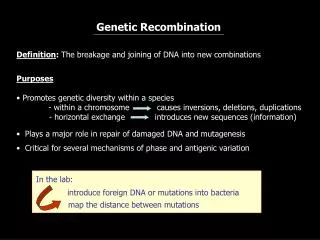
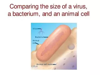
Genetic Recombination • If bacteria were incapable of genetic recombination, all members of a given species would be clones with differences arising only due to mutations in different lines • If bacteria could not share genetic information as other organisms do in sexual reproduction, there would be vastly less opportunity to adapt to different environments • As it turns out, bacteria are notorious for their ability to adapt; thus it is not surprising that elegant mechanisms exist for sharing genetic information
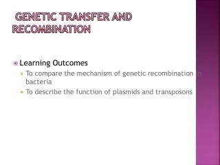
Ways Bacteria Exchange Genetic Material • Transformation - Bacteria take up DNA from their environment and incorporate it into their genome (i.e., the Griffith experiment) • Transduction - Movement of DNA between bacteria by viruses • Conjugation - The direct transfer of DNA by bacteria usually via plasmids
Conjugation Discovered by Joshua Lederberg and Edward Tatum in 1946. Unidirectional transfer of genetic material between donor and recipient bacteria cells by direct contact. Segment (rarely all) of the donor’s chromosome recombines with the homologous recipient chromosome. Recipients containing donor DNA are called transconjugants.
Fig. 15.2, Lederberg & Tatum (1946) Experiment demonstrating recombination in E. coli. Recombination of 2 complimentary auxotrophs gives rise to a strain that can synthesize all nutrients.
Fig. 15.3, Bernard Davis experiment demonstrated that physical contact is required for bacterial recombination.
Conjugation-transfer of the sex factor F: William Hayes (1953) demonstrated that genetic exchange in E. coli occurs in only one direction. Genetic transfer is mediated by sex factor F. Donor is F+ and recipient is F-. F is a self-replicating, circular DNA plasmid (1/40 the size of the main chromosome). F plasmid contains an origin sequence (O), which initiates DNA transfer. It also contains genes for hair-like cell surface (F-pili or sex-pili), which aid in contact between cells. No conjugation can occur between cells of the same mating type. Conjugation begins when the F plasmid is nicked at the origin, and a single strand is transferred using the rolling circle mechanism. When transfer is complete, both cells are F+ double-stranded.
Figs. 15.4 & 15.5a Transfer of the F factor
Conjugation of high-frequency recombinant strains: No chromosomal DNA is transferred by standard sex factor F. Transfer of chromosome DNA is facilitated by special strains of F+ integrated into the bacteria chromosome by crossing over. Hfr strains = high frequency recombination strains. Discovered by William Hayes and Luca Cavalli-Sforza. Hfr strains replicate F factor as part of their main chromosome. Conjugation in Hfr strains begins when F+ is nicked at the origin, and F+ and bacteria chromosomal DNA are transferred using the rolling circle mechanism. Complete F+ sequence (or complete chromosomal DNA) is rarely transferred (1/10,000) because bacteria separate randomly before DNA synthesis completes. Recombinants are produced by crossover of the recipient chromosome and donor DNA containing F+.
Fig. 15.5b Transfer of the HfrF+ factor
Fig. 15.6 Excision of the F+ factor also occurs spontaneously at low frequency. Begin with Hfr cell containing F+. Small section of host chromosome also may be excised, creating an F’ plasmid. F’plasmid is named for the gene it carries, e.g., F’ (lac)
Using conjugation to map bacterial genes: • Begin with two different Hfr strains selected from F+ x F-crosses and perform an interrupted mating experiment. • HfrH thr+ leu+ aziR tonR lac+ gal+ strR F-thr leu aziS tons lac gal strS • Mix 2 cell types in medium at 37°C. • Remove at experimental time points and agitate to separate conjugating pairs. • Analyze recombinants with selective media. • Order in which genes are transferred reflects linear sequence on chromosomes and time in media. • Frequency of recombinants declines as donor gene enters recipient later.
Fig. 15.7 Interrupted mating experiment
Fig. 15.7c, Genetic map-results of interrupted E. coli mating experiment.
Generating a map for all of E. coli: Location and orientation of the HfrF+ in the circular chromosome varies from strain to strain. Overlap in transfer maps from different strains allow generation of a complete chromosomal map. Fig. 15.8
Circular genetic map of E. coli Total map units = 100 minutes Time required for E. coli chromosome to replicate at 37°C.