Download
1 / 81
840 likes | 1.08k Vues
Explore fish kidney pathology focusing on haematopoiesis and host responses, introducing classes of pathogens affecting fish.

E N D
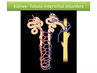
Systematic Fish Pathology: Part 2.Kidney I: Interstitial TissueGeneralised Responses Prepared by Judith Handlinger Fish Health Unit, Animal Health Laboratory, Department of Primary Industries & Water, Tasmania for The Australian Animal Pathology Standards (AAPSP) Program
Before Entering Training Program READ ME • This series of training modules has been prepared from a teaching slide set used for short courses on fish histopathology aimed at students with a range of prior knowledge of either pathology or fish. • Slides are representative of the pathology found in Australian fish in this (Tasmanian) fish laboratory, rather than a comprehensive record of all fish diseases, or diseases of all fish. There are some examples from other laboratories, including exotic diseases. • The major aim is to convey an approach to diagnosis, not to cover all fish diseases. The systematic approach is relevant, regardless of species (indeed this is just applying mammalian pathology training to fish). Nevertheless the pathology of any species can only be interpreted in comparison with the known normal. As there are more fish species than all the other vertebrates together, it is necessary to overcome the temptation to consider fish just a new “species” for study, and appreciate the diversity of fish species. • That this course does not cover all Australian fish, then, is within the context of using available material to impart a general knowledge of fish pathology including general patterns of fish diseases and an introduction to types of pathogen common in fish.
Acknowledgments. • Most material used has largely been generated within the Tasmanian Department of Primary Industries & Environment Fish Health Unit, and represents contribution of cases and photographs from many other contributors including Jeremy Carson, David Taylor, Stephen Pyecroft, Richmond Loh, Kevin Ellard, Paul Hardy-Smith, and Barry Munday. • Contributors of cases from other laboratories have been acknowledged wherever possible and specific material and photographs used with permission. Any inadvertent omissions in this regard are unintended. • Material exotic to Australia includes slides distributed for general teaching purposes and slides contributed specifically for the DPIW Fish Teaching set, and are acknowledged as such.
Introduction • Please refer to Part I “Consider the Fish” for an introduction to fish anatomy, histology and immunology, and how and why these differ from terrestrial animals. • I am starting the systematic examination of fish pathology with the kidney, because this is a major site of haematopoiesis, and a major reticulo-endothelial ¯ophage filtration bed. • Concentrating firstly on these interstitial elements provides a general introduction to host response cells and their reactions. The importance of this organ in generalised responses means this will also introduce most of the classes of pathogens affecting fish.
Kidney Anatomy The kidney is located in a retroperitoneal location adjacent to the dorsal margin of the abdominal cavity.
Margin of torn swim bladder Kidney Embryologically the head kidney is derived from the pronephros and consists mostly of haematopoietic tissue including lymphoid tissue. The posterior kidney (also called tail or body kidney) is derived from the mesonephros, and also contains the nephrons or tubular elements.
Micro-anatomy & histology The head kidney contains both haematopoietic tissue, and one of the major reticuloendothelial / macrophage filtration beds of the fish. The salmonids (particularly the Atlantic salmon) have been used as primary demonstration species. These are well studied, but regarded as a somewhat “primitive” teleost (bony fish) species, and major differences with “higher” species will be highlighted. The Goldfish is included as an example of the latter, together with other species. Remember these are included as representative examples, rather than as an indication of their importance. Considering the large range of fish species, familiarity with the normal of each species is essential to detect subtle changes but generalised knowledge is often all that is available.
Kidney anatomy Separate head and tail lobes are present in some fish, for example the Goldfish / carps. The separation is incomplete, with thin strands of interstitial tissue connecting the more obvious lobes.
The haematopoietic tissue is organised round a sinusoidal system of blood vessels that are lined by phagocytes. Normal juvenile Atlantic salmon. Ovary HeadKidney Dorsal skeletal muscle
Head kidney of a young Atlantic salmon showing sinusoid-like organisation of haematopoietic tissue. Note the regular pattern of heavily pigmented “melanomacrophages”, a typical end stage of fish phagocytosis. They are not melanocytes as such but contain mixed pigments which are mostly the end product of phagocytosis of pathogens or of host cells. The pigment may include melanin, haemosiderin, ceroid and lipofuscin. Note active haematopoietic tissue, separated by the slightly dilated vessels. Detail (another salmon) showing loose sinusoid-like organization Blood vessel
Detail of more mixed haematopoietic tissue, showing vascular and reticular framework, early development stages (probably red cell precursors – red arrows) & 2 melanomacrophages (white arrows).(Reminder – mature red cells are nucleated in all vertebrates except mammals.)
Another salmon with more lymphoid components and more mature polymorphonuclear granulocytes (arrows)
Head kidney with macrophages undertaking erythrophagy (the result of an ongoing toxicity, which we will come to later), & demonstrating the transition and darkening of the residual pigment through golden to black/brown.
Although melanomacrophages (MMs) show little aggregation in salmonids, which are regarded as a relatively “primitive” bony fish, and form at best a loose network round the margins of haematopoietic sinusoid groups, these cells typically actively migrate towards each other to form distinctive melanomacrophage centres (MMCs) in many fish species. Melanomacrophages and MMCs are also present in the spleen (another major filtration bed), and may be present in other tissues as an indicator of recent macrophage activity. MMCs are reported to play a significant role in antigen presentations to the lymphoid system, as well as such functions as preserving iron from effete erythrocytes for recycling.
Tail kidney of young Striped Trumpeter showing fewer but larger melanised bodies (melanomacrophage centres). This includes very dark MMCs & two paler centres (arrows). Macrophages have aggregated to form MMCs as their pigment is gradually transformed and condensed, so these represent more recent phagocytosis / tissue turnover. Individual macrophage shape is still visible in the young, but not the older MMCs
This shows the head kidney - tail kidney junction, separated by a blood vessel . Posterior kidney Details, showing glomeruli and the related convoluted tubules in tail kidney Blood vessel Note similar HP tissue in tail kidney, organised round the nephrons Head kidney
Tail kidney near junction showing extension of haematopoietic tissue between nephrons, and a similar distribution of melanomacrophages
Nephrons consist of a glomerulus and tubules, which are not oriented into a loop or specific pattern, but loosely convoluted near the glomerulus. • Consequently the kidney is not divided into cortex and medulla: • that was a consequence of the longer tubules needed for land animals for excreting nitrogenous products • fish excrete most nitrogenous by-products across the gills as ammonia. • the major function of the nephrons is water and bivalent ion control (and some toxin excretion). • Copious water is excreted by fish in fresh-water. Most water is resorbed by fish in marine environments. Glomerulus
In the Goldfish tail kidney MM centres are well developed, (white arrows) though the pigment is pale. (The pallor indicates residual pigments other than melanin.) Can you identify the bright eosinophilic bodies also present? (blue arrow).
Ah-Ha - ring-in!. These are thyroid follicles. Although the thyroid gland is usually in the expected location (between the lower jaws), individual follicles in other organs, especially this kidney are not unusual & in some fish are consistently present.
Thyroid gland in usual location– a loose collection of colloid containing follicles between the lower jaws of 5 gm Atlantic salmon.
The thyroid of this young salmon is forming a less diffuse organ.
The location of the normal (usual) thyroid can perhaps be better appreciated in this galaxid with goitre. (Yes, they do get goitre, particularly in freshwater from iodine deficient areas.) I have not recognized this in thyroid follicles within the kidney.
And another Ring-In: perivascular tissue in head kidney of an Angel Fish with skin lesions (ignore the necrosis). • What do you think this is? (Hint - mentally remove the haematopoietic tissue to long bones) Correct?You are left with cells round blood vessels anterior to the nephron-kidney that resemble those of the adrenal cortex and medulla - and are functionally the same. Termed inter-renal tissue (cortical equivalent) and chromaffin cells(medullary equivalent). Variations in the amount of inter-renal tissue may reflect area of section, physiological variations such as reproductive activity in some fish, or chronic stress. Note:frequently the distinction of these 2 cells types is not obvious in routine sections.
The effect of physiological state on the kidney interstitial tissue Changes in haematopoietic activityThis is often easier to assess in the tail or posterior kidney, where the nephrons help gage the amount of interstitial tissue present.
Posterior or tail kidney - normal young actively growing salmon with normal active haematopoiesis. … And that many of the haematic tissue cells how signs of maturation, such as a polymorphonuclear appearance. There are only occasional mitoses. Note the relative proportions of tubules and haematopoietic tissue
If we look at another area from the same kidney: Note occasional mitosis & mature cells e.g. granulocytes
Another example from this fish, illustrating that the distribution of interstitial tissue may not be uniform - need to consider the overview.
Haematopoietic Hyperplasia Increased haematopoietic activity in salmon smolt recently transferred to sea . Note high mitotic rate (arrows) and a dominance of immature cells including blast-like forms.
Hyperplasia is seen as an overall increase in interstitial cells seen both as high density - & slight separation of tubules. Mitoses ( ) Note both these animals are growing. Differences are only those of degree. “Smoltification” involves pre-adaptation to a marine environment. Animals entering the sea are readily able to regulate the high salt level and undergo a metabolic boost in thyroid & other hormones>increased growth>including increased HP activity.
Haematopoietic tissue depletion Haematopoietic tissue varies from sparse to normal, with some visible spaces (& a little tubule shrinkage artifact). In comparison, this salmon kidney shows closely packed tubules, with some spaces between. There is no evidence of cell loss due to cell death. Reduced interstitial tissue may reflect low levels of haematopoiesis (look for other indications of low growth & metabolic rate), but where there is an increase in tissue spaces this may also reflect mobilization of mature blood cells in response to stress or infections.
Another young salmon showing marked haematopoietic depletion, following (unidentified) stress. Only blood vessels separate many tubules.
And this one? No, not wildly depleted! This is normal. The fact that it has no haematopoietic tissue at all is the give-away. This is the tail kidney of a River Blackfish (Gadopsis marmoratus ) a species that has more complete separation of head kidney (haematopoietic tissue) and nephron containing tail kidney.
The effect of physiological state on the kidney interstitial tissue B. Changes in melanomacrophage activity.Increased melanomacrophage activity reflects phagocytic activity or tissue turnover. It may be accompanied by altered haematopoietic (HP) activity.For example, fish that are undergoing catabolism due to prolonged cessation of feeding will also show reduced HP activity.
Wild Brown trout: It is common to find more pigment in wild fish than similar aged farmed animals receiving a more concentrated & available diet & growing more rapidly. Judging whether this increase can be attributed to differences in diet or age, or reflect a pathological process of tissue turnover, is an interpretive skill requiring a good history to support histology findings.
Severe starvation Note that tubules are less prominent - urinary output is also reduced (marine, not eating, therefore minimal water intake with food.) “Failed smolt” some months after transfer to sea. Marked melanomacrophages increase is a reflection of cell turnover through catabolism. Starvation is common in young salmon (failed smolts) that were not adapted to a marine environment when transferred. To maintain salt balance by reducing seawater intake, they stop eating. Illustrates also the much more flexible food intake of fish: by this stage the fish had no fat, little muscle, but had been otherwise healthy, though close to death at this stage. Less affected fish would resume feeding if returned to freshwater.
Starvation, detail (x 40 objective). Note that in sufficient numbers, melanomacrophages will aggregate even in salmon. HP tissue is relatively sparse & most cells are mature, with negligible evidence of haematopoiesis –i.e. inactive.
Aggregation of melanomacrophages into MM centres is much stronger in “higher” fish species, such as this aged Orego Dory (Obx4)
Inflammation & degeneration Generalised interstitial reactions 1. Generalised bacterial infection patterns
Kidney from an Atlantic salmon with Yersinia rucheri septicaemia. What do you see? Correct? You see very little change, or possibly some reduction in interstitial tissue? This is not uncommon with acute septicaemia, although the kidney is a good site for isolation of bacteria due in part to the large vascular beds, and more florid lesions may be seen with heavy infection, later stages of infection, or highly toxic bacteria. Don’t be fooled by the apparent absence of bacteria. The paucity of interstitial tissue may reflect low background demand (slow growth etc), and / or mobilisation of leukocytes in response to the infection. This is a common pattern in the kidney with early septicaemias or with common secondary pathogens such as Aeromonas hydrophila.
The mobilisation reaction is not specific to septicaemia. This salmon shows more marked mobilisation in response to the stress of a jelly-fish sting. This will be reflected initially as a circulating leukocytosis. Note the spaces with little tubule shrinkage.
Another example of marked mobilisation, not associated with infection. Atlantic salmon recently transferred to sea. Cause of the moderate stress not identified.
The depletion in this kidney is also due to marked mobilisation of blood cells - this is due to septicaemia with Yersinia ruckeri.
Another subtle effect of septicaemia - slight congestion of the interstitial bed in a salmon with Vibrio anguillarum infection. (Vibrio bacteria are among the most common marine bacteria with a capacity for pathogenicity - that is, the ability to survive within the vertebrate host environment. This ability is reflected in the ability to grow on media such as blood agar, which will inhibits many aquatic bacteria.)
This is a kidney from another young Atlantic salmon with Y. ruckeri septicaemia. In this case the interstitial tissue is of moderate density, with no obvious depletion. & Pycnotic nuclear fragments are clearly visible (x 40 objective) At higher power the HP tissue does look less regular
(same kidney) Some areas show both pycnotic fragments and at 100x objective. …. … bacteria are visible within phagocytes ….. and blood vessels
Obviously the Y. ruckeri bacteria are in abundance, probably more so than in the previous example (where little was seen except interstitial depletion), though possibly also for a longer duration. Y. ruckeri are in unusual numbers in the next slide (different outbreak) Note: It has long been assumed that destruction of active tissue such as haematopoietic tissue is a direct effect of bacterial toxins. There has been discussion recently that bacterial diseases (of vertebrates generally) may also produce cell destruction via toxin “switching on” the programmed cell death or apoptosis, rather than direct damage of the toxin to the cell membrane etc. This does not produce the degree of host response to dead cell components that results from necrosis. Reference: Parrino J, Hotchkiss RS, Bray M. Prevention of immune cell apoptosis as potential therapeutic strategy for severe infections. 2007. Emerg. Infect Dis. 13(2):191-198. Available from http://www.cdc.gov/EID/content/13/2/191.htm (x100 Obj. Manipulated contrast to show bacteria)
Why so much detail on pycnosis (single cell death, contraction & fragmentation) with bacterial toxins? Bacterial are not the only (or even the most common), cause of HP necrosis. A good starting point for: Inflammation & degeneration Generalised interstitial reactions 2. Viral infections (& differential diagnosis)
This Redfin perch Perca fluviatilis(ASVP Slide of the Month, October 1988) also shows diffuse haematopoietic necrosis, which is typical of several of the most severe viral diseases of fish. This is an example of epizootic haematopoietic necrosis virus infection, causing the disease epizootic haematopoietic necrosis (EHN). In this case much of the HP tissue is intact, but pycnotic nuclear fragments are present. The pattern of necrosis is similar to the that of the previous slide (Y. ruckeri bacterial infection), which must therefore be considered under differential diagnosis. - and vice versa! It is common for bacterial infection to be secondary, so the absence of bacteria does not exclude viral infection. - in the previous case, samples from the salmon with Y. ruckeri were sent for virus isolation, with negative results. While HP necrosis is seen with overwhelming Y. ruckeri infection, this finding would always be considered with caution because of the need to exclude virus involvement.
(Same fish, liver): Although the kidney lesions were relatively mild, necrotic lesions (and some cytoplasmic inclusions) were also present in the liver, illustrating that although the HP tissue may be most heavily impacted, these viruses are systemic. The above virus (EHNV) is an iridovirus endemic in some parts of southern Australia, and can affect a number of species, including rainbow trout. Virus isolation on fish-specific cell lines is the preferred option for primary diagnosis, but appropriate cell lines are often not available. Those developed are often only useful for very specific viruses. However a suite of cell lines is available for most of the OIE listed haematopoietic viruses. Due to the systemic nature, the recommended range of tissues includes kidney, spleen (the other major reticular tissue / filtration organ of the fish), the liver, and for some viruses heart and / or encephalon. Serology is regarded as a poor test in fish as systemic antibody titres after infection are not reliably increased as titres may be influenced by variations in temperature or suppressed by stress. Thus there is an emphasis on developing tests such as PCR that are independent of the host response - though this is easiest to do once the virus has been isolated. Electron microscopy remains a valuable primary diagnostic tool.