Vertigo -BPPV
Vertigo -BPPV. W.M.C. Narampanawa. The Ear. Definition. An illusion or hallucination of movement which is usually rotational, either of oneself or the environment. Balance disorders. A common problem Many different potential etiologies Some time multifactorial

Vertigo -BPPV
E N D
Presentation Transcript
Vertigo -BPPV W.M.C. Narampanawa
Definition An illusion or hallucination of movement which is usually rotational, either of oneself or the environment
Balance disorders • A common problem • Many different potential etiologies • Some time multifactorial • Diagnostic & management challenge • Some time unable to make definitive diagnosis
Dizziness – four basic types • Vertigo – illusion of movements, usually rotational, can be an illusion of tilting or swaying • Pre syncope – light headedness • Disequilibrium – general sense of imbalance on walking • Others –(psycho physiologic) difficult to characterize
Introduction • Dizziness is a common presenting complaint • Dizziness may result from a disorder that affects any of the body parts involved in balance or from certain drugs. • The person's description of the problem and the results of a physical examination may suggest a cause, which may lead to additional tests.
vertigo • vertigo is the most common (40-50%) • Of the various causes of vertigo, benign positional vertigo (BPV) is the most common cause • Approximately 25-40% of patients who present with dizziness have BPV.
Near-syncope • Due to reduced blood flow to the entire brain and is classically described as feeling faint or lightheaded
Disequilibrium • Is essentially a gait disorder • Often caused by various neurological problems like cervical spondylosis, extra pyramidal disease and cerebellar disease • Patients typically describe their dizziness only when walking.
Psychophysiologic dizziness • This is the least understood and is thought to be due to altered central integration of sensory signals arising from normal end organs
BPPV/ BPV • BPV was first described by Adler in 1897 and then by Bárány in 1922 • Using positional testing, BPV can readily be diagnosed
BPPV • B” = Benign • Not a brain tumor • Can be severe and disabling
BPPV • “P” = Paroxysmal • Episodic, not persistent • Helpful feature in the differential diagnosis
BPPV • P” = Positional • Occurs with position of head • Turning over in bed • Looking up • Bending over
BPPV • V” = Vertigo • An illusion of motion • “The room is spinning” • Other descriptions • Rocking • Tilting • Descending in an elevator
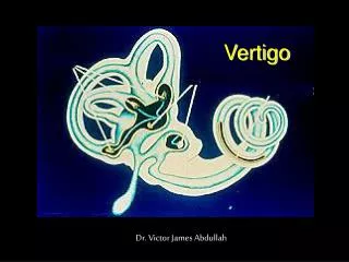
Anatomy: Utricle • Utricle • Connected to SCC • Contains endolymph • Otoliths (otoconia) • Calcium carbonate • Attached to hair cells • Macule (end organ)
Vestibular system • Tells brain which way the head moves without looking • SCC: angular acceleration • Utricle: linear acceleration
Pathophysiology of BPPV • Otoliths become detached from hair cells in utricle • Inappropriately enter the semicircular canal
Physiology • Normal situation • As one turns head to the right • Endolymph moves SCC receptors fire “head turning right” • Stop turning head endolymph stops moving SCC receptors stop firing “head has stopped moving”
Pathophysiology of BPPV • BPPV • Stop turning head otoliths keep moving drag endolymph receptors continue to fire inappropriately “head is still moving” • Eyes “head is NOT moving” • Brain room must be spinning in the opposite direction
Incidence • Incidence of BPV is 64 cases per 100,000 population per year (US) • Women are affected twice as often as men • in general, is a disease of elderly persons, although onset can occur at any age
Diagnosis - History • characteristically describe that the room or world is spinning • Diagnosis of posterior canal BPV is based on a characteristic history and a positive Hallpike test • Lateral Canal BPPV -Lateral Roll test
Vertigo may occur with • Rolling over in bed • Lying down • Sitting up • Leaning forward • Turning the head in a horizontal plane
Symptoms are usually worse in the morning • Nausea is typically present (vomiting is less common) • individual episodes of vertigo in BPV last for seconds at a time
Diagnosis - Examination • diagnosis of PC BPV is indicated by a positive Hallpike test • The neurologic examination is otherwise unremarkable
Dix - Hallpike test • Classic nystagmus occurs when the patient's head is dependent and turned to the affected side • The most common nystagmus seen is torsional or rotatory • Nystagmus usually occurs within 10 seconds after positioning but may present as late as 40 seconds
Dix – Hallpike test • Duration varies from a few seconds to a minute and associated the sensation of vertigo • Response fatigues if the patient is repeatedly placed into the provoking position
Hallpike test • Caution: For patients with cervical spondylosis • warn the patient that symptoms of vertigo • Instruct the patient to keep his or her eyes open no matter how bad he or she feels • Avoid in pts with IHD
Hallpike test • Seat the patient close enough to the end of the table • lies supine • head should be extended backward an additional 30-45°.
Hallpike test • Turn the patient's head 45° • This position orients the head such that the posterior semicircular canal is going to be in the same plane as the upcoming head movement
Hallpike test • lay the patient down until the head is dependent • This step does not need to be performed rapidly.
Hallpike test • Check for reproduction of symptoms and nystagmus • the fast phase of the nystagmus should be upbeat (toward the forehead)
Hallpike test • Return the patient to the upright position • Nystagmus may be observed in the opposite direction • Patient may describe that the world is spinning in the opposite direction • The neurologic examination findings should be otherwise normal; if not, strongly consider alternative diagnoses
Horizontal Canal BPV • Lateral Roll test Body supine Head inclined 30º Turn head to either side 2 variants Geotropic Apogeotropic
Geotropic LC BPPV • Free particles in the long arm of the LSC (Canalolithiasis) • Horizontal Ny. towards lowermost ear • Stronger Ny. on turning towards side of lesion.
Apogeotropic LSC BPPV • Cupololithiasis • Horizontal Ny.awayfrom lowermost ear • Stronger Ny. on turningaway from side of lesion.
Positional vertigo PERIPHERAL CENTRAL
Causes • Idiopathic (50-60%) • Infection (viral neuronitis) • Head trauma, especially in younger patients • Degeneration of the peripheral end organ • Surgical damage to the labyrinth
DD • Migraine • Labyrinthitis • Meniere’s disease • Vestibular neuronitis • Stroke • Acoustic schwanoma
DD • Chronic otomastoiditis • Medications (alcohol, phenytoin, diuretics, salicylates, quinidine, quinine, barbiturates, antibiotics) • Otosclerosis • Ototoxicity • Posttraumatic injuries • Vertebrobasilar insufficiency
Investigations • No pathognomonic laboratory test for BPV exists • Currently, no imaging study can demonstrate the presence of otoliths • Head CT scanning or MRI is indicated if the diagnosis is in doubt
Management • Epley maneuver • Medical treatment is generally ineffective but may be used to lessen the symptoms.
Epley maneuver Contraindications • Ongoing CNS disease (ie, stroke or transient ischemic attack [TIA]) • Unstable heart disease • Severe neck disease (eg, rheumatoid arthritis) or history of cervical spine fracture or surgery • Carotid bruit on examination indicating carotid stenosis • Body habitus preventing performance of the maneuver.
Epley maneuver • Goal is to move the otoliths out of the posterior semicircular canal and back into the utricle where they belong • The success rate of the Epley maneuver is very high (approximately 85-90%)
Epley maneuver general guidelines • The head must be in the dependent (head-hanging) position • Maintain each position until the symptoms and nystagmus have disappeared (at least 30 seconds) • If the patient cannot tolerate the maneuver because of vomiting or severity of the vertigo, premedicate with a vestibular sedative
Epley maneuver steps • patient sit upright on the bed with the head turned 45° to the affected side
Epley maneuver steps • Place your hands on either side of the patient's head and guide the patient down with the head dependent (as in the Hallpike test).
Epley maneuver steps • Rotate the head 90° to the opposite side with the patient's face upward and be sure to maintain the head-dependent position