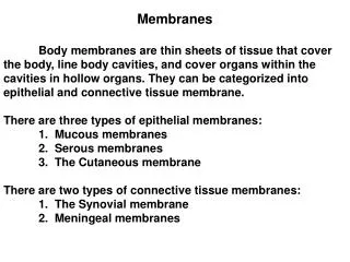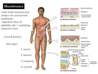
Function of Membranes
300 likes | 458 Vues
Function of Membranes. Compartmentalisation/ Dynamic boundary Isolation of cell from external world Alter cell form as necessary Division cell contents. Functions of Membranes. Selective permeability Control entry/exit in cells Control over organelle contents .Lysosome pH 5

Function of Membranes
E N D
Presentation Transcript
Function of Membranes Compartmentalisation/ Dynamic boundary • Isolation of cell from external world • Alter cell form as necessary • Division cell contents
Functions of Membranes • Selective permeability • Control entry/exit in cells • Control over organelle contents • .Lysosome pH 5 • Electrical isolation • Electrochemical gradient maintained • Neuronal function
Functions of Membranes • Localisation of Chemical reactions e.g. • Mitochondrial cristae - development of H+ ion gradients; required for ATP generation • Contain electron transport chain proteins • Chloroplast membranes - light gathering proteins • Lysosomes contain digestive enzymes
Functions of Membranes • Transport • Contain proteins for transport • Active transport processes • ATP dependent transport • co transporters • Pinocytosis, endocytosis • Including generation of gradients • Use of these gradients to provide energy for active transport
Functions of Membranes • Signal transduction • Receptors e.g. • Insulin receptor • Transduction mechanism • e.g. cAMP cascade requires enzymes adenylate cyclase
Functions of Membranes • Cell-cell recognition • glycoproteins • Cell-cell adhesion • Stick cells together
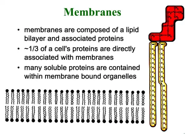
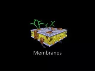
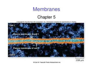
STRUCTURE • Phospholipid bilayer • FLUID MOSAIC MODEL • Proteins embedded in a sea of lipid • Cholseterol reduces fluidity • Proteins can be anchored by cytoskeleton • Proteins can span the membrane (transmembrane), • Extra/intracellular surface
REGULATION OF FLUIDITY Fatty acids are crucial regulators of fluidity – determined by chain length and degree of saturation Short chain fatty acids reduce the tendency of hydrocarbon chains to interact and hence increase fluidity The kinks in unsaturated fatty acids result in less stable van der Waals interactions with other lipids and hence increase fluidity High cholesterol content restricts the random movement of polar heads and decreases fluidity.
IMPAIRED FLUIDITY CAN DAMAGE CELLS Increased cholesterol content of red blood cell membranes is associated with severe liver disease eg. Cirrhosis Cholesterol content of red blood cell membranes is increased by 20-60%, leading to decreased fluidity Alters cell shape, impairs O2 transport, destruction of red blood cells and anaemia
PROTEINS • Key to (most) membrane functions are the proteins embedded/ attached in the membrane Pore forming proteins Carrier protein – active transport Receptor protein Membrane bound enzyme Cell-cell adhesion Cell-cell recognition Cytoskeleton
Channel Proteins Poison Dart Frogs- Batrachatoxin opens Na+ channels • Pore forming proteins • Transmembrane • Membrane spanning regions contain hydrophobic amino acids • Allow diffusion of polar molecules across cell membrane e.g. sodium ions • Na+ channel in cell membrane voltage gated (action potential) • Tend to show some selectivity • Water will also flow through a channel
Carrier Proteins • 3 methods • Facilitated diffusion – solute assisted by carrier protein to diffuse down concentration gradient, no additional energy supply needed. • Active transport - ATP hydrolysis provides the energy. e.g. Na+ K+ pump • Co transport of e.g. Na+ or H+ down their electrochemical gradient provides energy for a second solute to be moved against its concentration gradient e.g. Na+ are co transported with sugar molecules in the gut
Membrane bound enzyme • Galantamine (snowdrop), • Enzymes attached to the membrane • Can be external e.g. • cell wall synthesis in plants • Acetylcholinesterase (neuromuscular junctions) • Breaks down acetylcholine released by nerve cells to cause muscular contraction. • Poisoned by • Organophosphorous insecticides (OPs), • malathion (specific for insects - insecticide)
Poisoning causes muscle spasms, spasticity • Internally bound enzymes are often involvedin cell signalling cascades (see cell signalling section for details) • sarin (nerve gas),
Receptor Protein generating an intracellular response • Receptor protein specifically binds a ligand e.g histamine binds to a receptor on blood vessels. • Binding induces conformational change, on the receptor’s intracellular surface. • Variety of intracellular proteins activated • Production of second messengers or opening of an ion channel. • The second messengers (or ions) change cell function • e.g. Causes vasodilation/ increases permeability of blood vessel
Cell adhesion molecules • Responsible for connecting cells to cells • Often glycoproteins • Bind to proteins on neighbouring cells or extracellular matrix • Maintain tissue structure
Cell:cell recognition via a glycoprotein • Cell surface glycoproteins specific for each species • Determine e.g. blood group (causes difficulty for blood transplants/ xenografts)
Membrane protein attached to the cytoskeleton • Membranes are fluid and proteins can move around in them • But – some proteins may need to be localised e.g. • Underneath a synaptic cleft or neuromuscular junction • Specific localisation on cells to give sidedness • e.g. intestinal lumen • Cytoskeletal proteins can anchor membrane proteins in a specific location
Cytoskeleton (a) Light micrograph of the cytoskeleton. The microtubles are green and the microfilaments are blue. Intermediate filaments form most of the rest of the network. (b) Scanning electron micrograph of a portion of a cell's cytoskeleton. • Intricate network of thread-like filaments • Microfilaments (or Actin filaments) • Intermediate filaments • Microtubules • Support the interior, produce movements, shape changes
Microfilaments • Actin filaments (microfilaments) • Two actin strands twisted together • “rope” approx. 7nm diameter • Concentrated under the cell membrane • Important for cell movements • Dynamic
Intermediate filaments • Provide mechanical strength (important for animal cells) • Act like a scaffold
Microtubules • Hollow tubes made from tubulin • Heterodimers (one and one ) arrange to form 13 protofilaments • Important in cell division forming spindle fibre • Also involved in intracellular transport
Centrosome (Microtubule organising Centre) • Area in the cell which controls polymerisation, depolymerisation of microtubules • Centrioles are found there (animal cells only) • Plant cells have MOC, but no centrioles
CytoskeletonCytoskeleton and its major functionsProvides internal support and streanghtDifferent types of fibers all supply the same need The life a cytoskeleon is never complete. Its chief functions include movement for the cell, movement of material through the cell, maintaining the shape of the cell. But keeping the cell from getting smashed by other cells and moving the cell to where it need to be are its main roles. Early on, when not much was known about the structure of the cell, scientist believed that the cytoplasm that surrounds the organelle was completely random. As the years went on, better microscopes were invented, and better techniques were improved upon for making a slide, "a lacy network of fibers was revealed." These fibers look similar to girders that hold up a bridge, so it was hypothesized that they would do the same for the cell, hold its shape. These fibers can be broken down into three main groups: Microfilaments, microtubules, and intermediate filaments, all of which can be recognized by there structure and their protein makeup. Regardless of their differences all three of them serve the same goal in the cell, to make the cell more ridged.MicrofilamentsThey, along with other proteins and ions, are responsible for every muscle contraction The microtubules and the microfilaments play a role in whole cell activities, including cell division, contraction of cell, and the crawling of the cell to a new location. Also they help in movement of vesicles, small sacs used for holding molecules, within the cell. The microfilaments resemble a string of beads of the protein actin, thus earning them the name of filaments. These filaments are the smallest cytoskeletal component, ranging in at 6 nanometers. These actin proteins play a large role in muscle contraction. A model was made by Hugh Huxley that shows that the actin proteins are in alternating rows that alternate with myosin. The contraction of the muscle is caused by a calcium atom that excites the myosin, thus grabbing the actin and causing the muscle to shorten. After more studying of this event, it was learned that ATP was required to grab the actin.MicrotubulesThese together wiht centrioles aid in cell division by pulling apart chromosomesMovement is a result of the breaking down and building up of these internal stuctors of the cytoskeleton The thickest the components are the microtubules, at 22 nanometers. These were noticed to be in the cell since the 1950’s but could not be studied until 1963. Each tube is filled with the protein tubulins. With this protein, the microtubules can shrink and grow. One of the most important jobs of these tubes is to aid in cell division: It releases centrioles, which or composed of microtubules, which migrate to the cells poles. After doing this the chromosomes as move to the poles as result of the centrioles, this proves that cell division does not require ATP. When the cell is not dividing the microtubules carry vesicles to new locations. The ability of the microtubules and microfilaments to breakdown and build up aid in the movement of a cell. Although there is the intermediate filaments that are very stable. They play important roles with cells that do mechanical stress. Research is being done to see whether these also play a role in cancerous cells. As we have just learned, the cytoskeleton plays a major role in almost every function of the cell.
The Cytoskeleton • Every cell contains specialized cytoplasmic proteins which serve as a support and contractile system, maintaining or changing cell shape. The cytoskeletal structures include the microtubules, microfilaments, and intermediate filaments. • Microtubules • Cytoplasmic microtubules appear as 25 nanometer tubular structures and are readily assembled and disassembled from cytoplasmic pools of the protein tubulin. Microtubules are fairly rigid and play a role in the maintenance of cell shape (Microtubules 1). They are associated with cilia formation (from basal body) (Microtubules 2), and spindle apparatus formation (from the centriole) during cell division (Microtubules 3). • Microfilaments • Microfilaments are fine, thread like structures about 6 to 7 nanometers in diameter. An analogy sometimes employed to indicate function is that the microtubules act as the "bones" of the cell, whereas the microfilaments act as the "muscles" since they provide for movement and shape change. Microfilaments are composed of the contractile protein actin and represent a primitive contractile system, forming large bundles called stress fibers (Microfilaments 1). In most cells, the microfilaments are found in a band just under the plasma membrane (Microfilaments 2). • Intermediate Filaments • Intermediate filaments are generally 8 to 12 nanometers in diameter and biochemically are a heterogeneous group (keratan, dermatan, desmin, and vimentin are a few of the biochemical species of intermediate filaments). The intermediate filaments are not contractile and serve exclusively in a supportive role. They are frequently grouped into delicate bundles (fibrils) in the cytoplasm, suited to meet stress and provide an overall girder-like support system.