
Neuroprotective Effects of Statins in Ischemic Retinal Injury
This study explores the potential neuroprotective role of statins in retinal ischemic-reperfusion injury, investigating how simvastatin treatment affects retinal function post-injury in an experimental glaucoma model.

Neuroprotective Effects of Statins in Ischemic Retinal Injury
E N D
Presentation Transcript
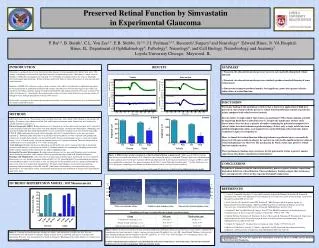
1500 1500 1000 1000 500 500 0 0 INTRODUCTION -500 -500 -1000 -1000 Glaucoma is a group of diseases that share in common damage to retinal ganglion cells and the optic nerve. The damage is frequently associated with significant increases in intraocular pressure. Glaucoma is a major cause of blindness worldwide accounting for an estimated 15 % of blindness. Ischemic insult to the retina is frequently observed in open angle glaucoma. Retinal ischemic insult often leads to serious disturbances of neuronal and glial retinal elements. Inhibitors of HMG-CoA reductase, such as statins, continue to be widely used clinically as lipid-lowering agents for the management of hypercholesterolemia and coronary atherosclerosis. Recent data suggests that statins can ameliorate neurologic ischemic damage by improving blood flow and making neuronal tissue more resistant to the effects of ischemia (1). Specifically, the neuroprotective effects of statins in the brain after an ischemic stroke are thought to occur through upregulation of brain eNOS activity. In this study we propose to determine the neuroprotective effects of statins on retinal tissue in an established model of ischemia-reperfusion injury. -1500 -1500 1500 1500 Vehicle Simvastatin 1000 1000 500 500 Pre-ischemia Pre-ischemia 0 0 Amplitude (µV) Amplitude (µV) -500 -500 -1000 -1000 -1500 -1500 Post-ischemia Post-ischemia Amplitude (µV) Amplitude (µV) Scotopic +5 dB Flash Scotopic +5 dB Flash 50 50 -150 -150 -100 -100 -50 -50 0 0 100 100 150 150 200 200 250 250 300 300 Time (ms) Time (ms) a-wave b-wave DISCUSSION SUMMARY CONCLUSIONS Prophylactic administration of simvastatin modestly protects against ischemic-reperfusion dependent deficits in retinal function. These preliminary findings suggest that statins may have neuroprotective effects in this experimental model of glaucoma. • Transient elevation of intraocular pressure in Lewis rats markedly diminished retinal function • Transient elevation of intraocular pressure similarly produced marked thinning of inner retinal layers • Simvastatin treatment produced modest, but significant, protection against ischemic-induced loss of retinal function The major finding of this preliminary study is that a short-term application of high-dose parenteral simvastatin modestly preserves retinal function following ischemic-reperfusion injury compared with vehicle-treated controls. Recent studies strongly suggest that statins can ameliorate CNS ischemic damage, possibly by improving blood flow to affected tissue (5). Despite the significance of these early observations, there has been a paucity of studies examining the potential neuroprotective role of statins in retinal ischemic-reperfusion injury. In fact, only a single article has been published implicating statins as neuroprotective agents following retinal ischemic injury secondary to optic nerve ligation (6). Here, we found that retinal function following ischemic-reperfusion injury was modestly preserved with simvastatin treatment. In contrast, no statistically significant preservation of retinal morphology was observed. The mechanism by which statins may preserve retinal function remains unclear. These preliminary findings raise awareness for the potential of statins to protect against some forms of ischemic-reperfusion retinal injuries. OS OS IS IS METHODS ONL ONL INL INL Adult male Lewis rats (n = 9 per group) were treated for three days with vehicle (10% ethanol) or simvastatin (20 mg/kg). Treatments were administered 48h, 24h, and 2h prior to ischemic insult. Simvastatin was activated by alkaline hydrolysis prior to use. Electroretinography (ERG):Retinal function was evaluated in rats before simvastatin treatment and 1 week after retinal ischemia-reperfusion injury. Rats were dark-adapted overnight, anesthetized with ketamine (100mg/kg) and xylazine (5mg/kg) and their pupils dilated. ERG’s were recorded with a stainless steel wire electrode loop (LKC UTAS-E 3000) contacting the corneal surface through a layer of 1% methylcellulose, as we have previously reported (2). Platinum needle electrodes were placed in the cheek and tail served as reference and ground leads, respectively. A single-flash stimulus was presented in order of increasing luminance across a 4-log unit stimulus range (-3.8 to +1 log cd sec/m2) with an interstimulus interval of 1 minute in duration. The body temperature of anesthetized rats was maintained at 37 ºC with a heating pad. Acute Ischemia: Retinal ischemia was induced in anesthetized Lewis rats by transient elevation of intraocular pressure for 60 minutes as previously described (3). Retinal ischemia was confirmed by blanching of the ocular fundus and the collapse of the retinal artery by indirect ophthalmoscopy. Intraocular Pressure Measurement: Baselineintraocular pressure was measured 5 minutes prior to ischemic insult, during the procedure, and 5 minutes following reperfusion using a hand-held TonoLab tonometer. Histology and Morphometry: One week after transient retinal ischemic injury anesthetized rats were sacrificed. Enucleated eyes were fixed in phosphate-buffered (pH 7.4) 2.5% glutaraldehyde-2% paraformaldehyde solution and embedded in epoxy resin. Sections (1 µm), cut along the vertical meridian of the eye and passed through the optic nerve head, were stained with toluidine blue. The inner retinal thickness (between the inner limiting membrane and the boundary of the ONL and OPL) and whole retinal thickness were measured as previously described (4). For each retinal section, four separate measurements were performed at 365μm and 913μm from the optic nerve head. IPL IPL GCL REFERENCES GCL 1. Sironi L, Cimino M, Guerrini U, Calvio AM, Lodetti B, Asdente M, Balduini W, Paoletti R, Tremoli E. Treatment with statins after induction of focal ischemia in rats reduces the extent of brain damage. Arterioscler Thromb Vasc Biol 2003;23:322-327. 2. Bu P, Von Zee CL, Basith B, Stubbs EB, Perlman JI.ERG Changes and D-Aspartate Uptake in Experimental Diabetic Retinopathy. 2006 Annual Meeting Abstract and Program Planner accessed at www.arvo.org. Association for Research in Vision and Ophthalmology. Abstract 4242. 3. Perlman JI, McCole SM, Pulluru P, Chang J, Lam TT, Tao MOM. Disturbances in the distribution of neurotransmitters in the rat retina after ischemia. Cur Eye Res 1996;15: 589 – 596. 4. Shibuki H, Katai N, Kuroiwa S, Kurokawa T, Arai J, Matsumoto K, Nakamura T, Yoshimura N. Expression and neuroprotective effect of hepatocyte growth factor in retinal ischemia-reperfusion injury. Invest Ophthalmol Vis Sci 2002;43:528-536. 5. Delanty N, Vaughan CJ. Vascular effects of statins in stroke. Stroke. 1997;332:481-487. 6. McGwin G, McNeal S, Owsley C, Girkin C, Epstein D, Lee PP. Statin and other cholesterol-lowing medications and the presence of glaucoma. Arch Ophthalmol. 2004;122: 822-826. 50 µm Naïve, non-ischemic retina Vehicle-treated ischemic retina Simvastatin-treated ischemic retina Preserved Retinal Function by Simvastatin in Experimental Glaucoma P. Bu1,4, B. Basith1, C.L. Von Zee1,7, E.B. Stubbs, Jr.3,6, J.I. Perlman2,4,5, Research1,Surgery2 and Neurology3 Edward Hines, Jr. VA Hospital, Hines, IL. Department of Ophthalmology4, Pathology5, Neurology6, and Cell Biology, Neurobiology and Anatomy7 Loyola University Chicago, Maywood, IL RESULTS Figure 2.Effect of simvastatin-treatment on retinal function following ischemic insult. (Upper panels) ERG recordings from Lewis rats treated with vehicle (10 % ethanol) or simvastatin (20 mg/kg) as indicated. Tracings shown were obtained prior to (pre-ischemia) and 1-week (post-ischemia) following ischemic insult. (Lower panels) Quantitative changes in ERG a- and b-wave amplitudes as indicated. Pre, prior to ischemic insult; Post, 1-week following ischemic insult. * p<0.05, Student’s t-test (n = 8 per group) comparing post-ischemic insult data between vehicle- and simvastatin-treated rats. Short-term parenteral administration of simvastatin modestly preserves retinal function following ischemic insult. ISCHEMIC-REPERFUSION MODEL: IOP Measurements Figure 3. Effect of simvastatin on preservation of retinal morphology following ischemic insult. Shown are representative toluidine blue-stained retinal sections from naïve, vehicle-, and simvastatin-treated (20 mg/kg) Lewis rats. IRL, Inner Retinal Thickness (between the inner limiting membrane and the boundary of the ONL and OPL). *p < 0.02, **p < 0.003 Student’s t-test comparing vehicle- and simvastatin-treated groups to non-ischemic controls. Ischemic-reperfusion injury produced a marked thinning of the inner retinal layers and whole retina. Simvastatin-treatment modestly preserved retinal morphology following ischemic insult. Figure 1. Transient intraocular pressure changes in vehicle- and simvastatin-treated Lewis rats. Pressure measurements were taken prior to ischemia, during ischemic insult as indicated, and again at 5 minutes post-reperfusion. Pressures were measured with a TonoLab tonometer. Blanching of the ocular fundus was evident in all cases at onset of ischemia. Acknowledgements: This work was supported by Department of Veterans Affairs (C3638R) and The Richard A. Peritt Charitable Foundation.


