IRON
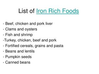
IRON. 7 mg/1000 cal in diet; 10% absorbed Heme iron absorbed best, Fe 2+ much better than Fe 3+ Some foods, drugs enhance and some inhibit absorption of ionic iron Ability to regulate absorption limited Absorption in proximal small intestine

IRON
E N D
Presentation Transcript
IRON • 7 mg/1000 cal in diet; 10% absorbed • Heme iron absorbed best, Fe2+ much better than Fe3+ • Some foods, drugs enhance and some inhibit absorption of ionic iron • Ability to regulate absorption limited • Absorption in proximal small intestine • Absorption greater if there is erythroid hyperplasia or hypoxia
Regulation of iron uptake and storage. Hepcidin, a peptide produced in the liver, is a key regulator of iron release from villus enterocytes and macrophages. Hepcidin, whose production is upregulated by high plasma iron levels or inflammation, inhibits iron release from these cells and lowers GI absorption. Low iron levels decrease hepcidin production, which in turn stimulates iron absorption and release into the blood. The HFE gene modulates hepcidin production. Mutations in HFE can cause diminished hepcidin release, and can eventually cause iron overload (hereditary hemochromatosis). N Engl J Med 2004;350:2383
1 cc of red cells contains about 1 mg iron 1 cc of whole blood contains 0.5 mg iron
IRON BALANCE • 1-2 mg/day lost via desquamation, GI blood loss in adult • Negative iron balance possible in early childhood • Menstruation, pregnancy, lactation promote negative balance • Positive balance (and eventual iron overload) can occur in inherited disorders (hemochromatosis), or as a result of repeated blood transfusions
♀ ♂
Plasma levels of iron are closely regulated to ensure a daily supply of about 20 mg to the bone marrow for incorporation into hemoglobin. Most of the iron in the plasma derives from the continuous breakdown of hemoglobin in senescent red cells by macrophages. 1 to 2 mg per day of iron is also taken up by duodenal enterocytes and transferred to the plasma compartment or, depending on body needs, stored in the enterocytes as ferritin. These stores are eliminated when enterocytes are sloughed at the end of their life cycles; apart from menstrual blood loss, this is the only significant means by which excess body iron is excreted. Iron recycled by macrophages and that absorbed from the gut is loaded onto serum transferrin and delivered primarily to the bone marrow for reincorporation into new red-cell precursors. The remaining body iron (about 1000 mg) is stored, primarily in hepatocytes. N Engl J Med 2004;350:2383
IRON TRANSPORT AND STORAGE • Absorbed iron oxidized to Fe3+ form • Bound tightly to transferrin in blood • Transferrin = total iron binding capacity (TIBC) • Normally serum iron about 100 μg/dl, TIBC about 300 μg/dl • Iron transferred to cells and reduced to Fe2+ form, then inserted into heme or stored • Storage iron (Fe3+) bound to ferritin • Hemosiderin is denatured ferritin • Small amount of ferritin in blood (nanograms)
ASSESSMENT OF BODY IRON • Serum iron low in iron deficiency, inflammation • TIBC high in iron deficiency, normal or low in inflammation • Ferritin low in iron deficiency, increases in inflammation • Marrow iron stores absent in iron deficiency • Combination of iron deficiency and inflammation makes assessing iron stores difficult
TESTS OF IRON STATUSPractical aspects • Low serum ferritin almost always indicates iron deficiency • Low serum iron and high TIBC almost always indicate iron deficiency • Ferritin > 100 rarely found in iron deficiency • Exception - liver inflammation/necrosis • Normal serum iron rarely found in iron deficiency • Exception - iron deficiency recently treated with oral iron • When TIBC is low or normal, low serum iron not a reliable indicator of iron deficiency • Iron deficiency may be hard to diagnose via blood tests in setting of inflammation (eg, low iron, low TIBC, intermediate ferritin level) • Therapeutic trial of iron +/- EPO a reasonable alternative to marrow biopsy
HYPOPROLIFERATIVE ANEMIA Lower than expected red cell production (erythroid cells in marrow and reticulocytes) for degree of anemia
HYPOPROLIFERATIVE ANEMIACauses and examples • Insufficient iron for hemoglobin synthesis • Iron deficiency • Diminished iron release from storage sites • Inflammation • Blunted or impaired erythropoietin production • Inflammation, renal failure • Inhibition of red cell progenitor proliferation by cytokines • Inflammation • Marrow damage • Aplastic anemia, myelophthisic disease
IRON DEFICIENCY • Most common cause of anemia • Usually due to chronic blood loss • Exceptions: rapidly growing child, malabsorption • In young women this is usually due to menstrual blood loss pregnancy • In anyone else must rule out GI blood loss • Esophageal disease, hiatal hernia, ulcer, inflammatory bowel disease, angiodysplasia, hemorrhoids, cancer
Iron deficiency Atrophic glossitis “Spoon nails”
Absent iron stores Normal iron stores
IRON DEFICIENCY ANEMIATreatment • Oral ferrous salts • Some patients have GI side effects • Oral iron-polysaccharide complex • IV iron dextran • If oral iron not absorbed or not tolerated • Slight risk of anaphylaxis • Should see increased hemoglobin within 2-3 weeks
IRON OVERLOAD • Hereditary hemochromatosis • Autosomal recessive, HFE gene; genotype common but low penetrance • Other inherited disorders • Mutations in other genes that regulate iron metabolism • Africans, African-Americans • Chronic ineffective erythropoiesis • Thalassemia • Repeated transfusion • Toxicity after about 100 Units
IRON OVERLOAD • Increased serum iron and high transferrin saturation (90%+ in hemochromatosis) • Very high serum ferritin • Increased liver and marrow iron • Quantitation of liver iron best indicator of severity • DNA test available for hereditary HC
IRON OVERLOADClinical consequences • Cirrhosis, hepatocellular carcinoma • Cardiomyopathy, heart failure • Endocrine failure (esp diabetes) • Arthropathy • Treatment of hereditary HC by phlebotomy prevents these problems and can reverse early tissue damage