Download
1 / 65
700 likes | 1.19k Vues
Diseases of the oesophagus. Oesophageal motility disorders. Key facts A spectrum of diseases involving failure of coordination or contraction of the oesophagus and its related muscular structures. Pathological features

E N D
Oesophagealmotilitydisorders Key facts • A spectrum of diseases involving failure of coordination or contraction of the oesophagus and its related muscular structures. Pathological features • In some cases degeneration of the inner and outer myenteric plexuses can be demonstrated but often no structural abnormality is seen.
Clinical features • Achalasia • Peak ages of incidence in young adulthood (idiopathic) and old age (mostly degenerational). • Slowly progressive dysphagia: initially worse for fluids than solids. • Frequent regurgitation of undigested food common late in the disease. • Secondary recurrent respiratory infections due to aspiration.
Diffuse oesophageal spasm • Commonest in young adults; • Characterized by acute pain along the length of the oesophagus induced by ingestion, especially of hot or cold substances (odynophagia).
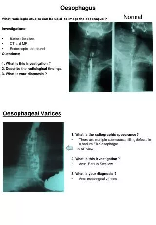
Diagnosis and investigations • Achalasia • Video bariumswallow. A characteristicfailureofrelaxationoftheloweroesophaguswithasmoothoutline rat'stail or birdbeak. • Oesophagealmanometry. Hypertonicloweroesophagealhighpressurezonewithfailureofrelaxationnormallyinduced by swallowing. In chroniccasestheproximaloesophagusmaybeadynamic. • Oesophagoscopy. To excludebenign and malignantstrictures.
Diagnosis and investigations • Diffuseoesophagealspasm • Video bariumswallow. Corkscrewappearanceoftheoesophaguscaused by dyscoordinateddiffusecontractions. • Oesophagealmanometry. Diffusehypertonicity and failureofrelaxation. Little or no evidenceofcoordinatedprogressiveperistalsisduringepsiodesbutnormalperistalsiswhenasymptomatic. • Oesophagoscopy. Required to excludeunderlyingassociatedmalignancy.
Treatment • Achalasia • Endoscopically guided controlled balloon dilatation (fixed pressure)successful in up to 80% of patients. Low complications rate (perforation). May need multiple procedures over time. • Botulinum toxin injections: success in some patients failing dilatation. • Surgical myotomy (Heller's cardiomyotomy). Open or thoracoscopically performed division of the lower oesophageal muscle fibres. Highly successful in resistant cases. Most applicable to young patients. • Specific complications include reflux, obstruction of gastro-oesophageal junction, oesophageal perforation.
Diffuseoesophagealspasm Oral calciumchannelblockers, or relaxants, e.g. benzodiazepines. Long-actingnitric oxide donors (smoothmusclerelaxant). Widespreadoesophagealpneumaticdilatations (oftenrepeated). Longsurgicalopenmyotomyrarelyundertaken.
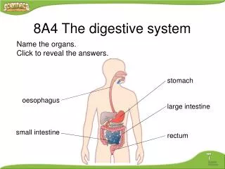
Keyrevisionpointsanatomy and physiologyoftheoesophagus Upper 2/3. Stratifiedsquamousepithelial-lined (developssquamouscarcinoma), striatedskeletalmuscle, lymphaticdrainage to neck and mediastinalnodes, somaticinnervationofsensation (e.g. moderatelyaccuratelocationoflevelofpathology). Lower 1/3. Transition to columnarepithelium (developsadenocarcinoma), transition to smoothmuscle, lymphaticdrainage to gastric and para-aorticnodes, visceralinnervation (poor localizationofpathology).
Keyrevisionpointsanatomy and physiologyoftheoesophagus Gastro-oesophagealjunctionis site ofporto-systemicanastomosis (betweenleftgastric and (hemi)azygousveins) maydevelopgastric or oesophagealvarices. Upperoesophagealsphincter (UOS) = cricopharyngeus. Loweroesophagealsphincter (LOS) = functionalzoneofhighpressureabovethegastro-oesophagealjunction. Relaxantsincludealcohol. Swallowingrequiresintact and coordinatedinnervationfromvagus (UOS, oesophagus, LOS) and intramuralmyentericplexus.
Pharyngeal pouch – Zenker'sdiverticulum Key facts • An acquired pulsiondiverticulum arising in the relatively fibrous tissue between the inferior constrictor and cricopharyngeus muscle: Killian's dehiscence. • Arises primarily as a result of failure of appropriate coordinated relaxation of the cricopharyngeus causing increased pressure on the tissues directly above during swallowing. • Typically occurs in the elderly.
Zenker'sdiverticulum Key facts • Associated with lower cranial nerve dysfunction (e.g. motor neuron disease, previous CVA). Pathological features • Acquired diverticulum: fibrous tissue and serosa without muscle fibres in most of the wall. • Tends to lie to one side of the midline due to the cervical spine directly behind.
Zenker'sdiverticulum Clinical features • Upper cervical dysphagia. • Intermittent lump appearing to the side of the neck on swallowing. • Regurgitation of foodundigested.
Zenker´sdiverticulum Diagnosis and investigations • Diagnosis may be made on observed swallowing with a transient neck swelling appearing. • Video barium swallow will show filling of pouch. • Gastroscopy should be avoided unless there is a question of associated pathology since the pouch is easily missed and easily damaged or perforated by inadvertent intubation.
Zenker´sdiverticulum Treatment • Endoscopic stapled pharyngoplasty: side to side stapling of pouch to the upper oesophagus, which also divides the cricopharyngeus muscle
Hiatus hernia Key facts • The presence of part or all of the stomach within the thoracic cavity, usually by protrusion through the oesophageal hiatus in the diaphragm • Very common; majority are asymptomatic. • May or may not be associated with gastro-oesophageal reflux disease. • Predisposing factors: obesity, previous surgery.
Hiatushernia Clinico-pathological features • Sliding hernia • Results from axial displacement of upper stomach through the oesophageal hiatus, usually with stretching of the phrenico-oesophageal membrane. • By far the commonest form. May result in GORD.
Hiatushernia Clinico-pathological features • Rolling (paraoesophageal) hernia • Results from the displacement of part or all of the fundus and body of the stomach through a defect in the phrenico-oesophageal membrane such that it comes to lie alongside the normal oesophagus. • Much less common. • Symptoms include hiccough, pressure in the chest, odynophagia. • May result in volvulus or become incarcerated and cause obstruction.
Diagnosis and investigations • Video barium swallow usually identifies the type and extent. • CT scanning of the thorax is the investigation of choice in acute presentations.
Treatment • Medical (mainlyfor GORD symptoms) • Reduceacidproduction. Stop smoking, loseweight, reducealcoholconsumption. • Counteractacidsecretion: protonpumpinhibitors, symptomaticreliefwithantacids. • Promoteoesophagealemptying : promotilants, e.g. metoclopramide.
Surgical • Rarely required. Indicated for: • persistent symptoms despite maximal medical therapy; • established complications of rolling hernia such as volvulus or obstruction. Elective procedure of choice is open or laparoscopic reduction of the hernia and fixation (gastropexy), usually with plication of the oesophageal opening (curalplication), occasionally with a fundoplication (e.g. Nissen's operation) if GORD symptoms predominate. Acute presentations may require a partial gastrectomy.
Gastro-oesophagealrefluxdisease GORD Key facts • Pathologically excessive entry of gastric contents into the oesophagus. • Reflux occurs in normals up to 5% of the time. • Commonest in middle-aged adults. • Usually due to gastric acid but also due to bile reflux. • Contributory factors include: • reduced tone in the lower oesophageal sphincter: idiopathic, alcohol, drugs, previous surgery, secondary to existing peptic stricture. • increased intragastric pressure: coughing, delayed gastric emptying, large meal.
Gastro-oesophagealrefluxdisease GORD Pathological features • Oesophagitis • Results in inflammatory changes in the squamous lined oesophagus. • Varies in severity from minor mucosal erythema and erosions to extensive circumferential ulceration and stricturing. (graded I to IV).
Gastro-oesophagealrefluxdisease GORD Pathological features • Stricture • Chronic fibrosis and epithelial destruction may result in stricturing. • Eventually shortening and narrowing of the lower oesophagus. • May lead to fixation and susceptibility to further reflux.
Gastro-oesophagealrefluxdisease GORD Clinical features • Dyspepsia may be the only feature; may radiate to back and left neck. • True reflux may occur with acid in the pharynx. • Commonly worse at night, after large meals, and when recumbent. • Dysphagia may occur if there is associated ulceration or a stricture.
Gastro-oesophagealrefluxdisease GORD Pathological features • Oesophagealmetaplasia Barrett'soesophagus • Maydevelopas a resultofgastro-oesophagealreflux; possibly more commonly in biliaryreflux. • Normalsquamousepitheliumisreplaced by columnarepithelium. • Dysplasia and premalignantchange (metaplasia) mayoccur in thecolumnarepithelium.
Gastro-oesophagealrefluxdisease GORD Diagnosis and investigations • Under the age of 45 • Symptoms are relatively common and can be treated empirically. Investigation is only required if symptoms fail to respond to treatment. • Over the age of 45 • Reflux can be confirmed by 24h continuous pH monitoring. Peaks of pH change must correspond to symptoms. • Endoscopy should be performed in all new cases over the age of 45 to exclude oesophageal malignancy.
Gastro-oesophagealrefluxdisease GORD Treatment • Medical • Reduceacidproduction: smoking, weight, alcoholconsumption. • Counteractacidsecretion: protonpumpinhibitors (e.g. omeprazole 20mg od), symptomaticreliefwithantacids (e.g. Gaviscon 10mL PO od). • oesophagealemptying: promotilants, e.g. metoclopramide 10mg tds PO.
Gastro-oesophagealrefluxdisease GORD • Surgical • Procedureofchoiceislaparoscopicfundoplication, Nissen'soperation (wrappingfundusofthestomacharoundtheintraabdominaloesophagus to augmenthighpressurezone).
Gastro-oesophagealrefluxdisease GORD • Surgical • Rarelyrequired. Indicatedfor: • persistentsymptomsdespitemaximalmedicaltherapy; • largevolumerefluxwith risk ofaspirationpneumonia; • complicationsofrefluxincludingstricture and severe ulceration. • Uncertain role in thepreventionofprogressivedysplasiainBarrett'soesophagealmetaplasiaintheabsenceofsymptoms.
Oesophagealtumours Keyfacts and pathologicalfeatures • There are severaltypesofoesophagealtumours. • Adenocarcinoma • Rapidlyincreasingincidence in Western world: 5:1 (M:F) • Commonest in Japan, northernChina, and SouthAfrica, • Associatedwithdietarynitrosamines, GORD, and Barrett'smetaplasia. • Typicallyoccurs in thelowerhalfoftheoesophagus.
Oesophagealtumours Keyfacts and pathologicalfeatures • Squamouscarcinoma • Incidenceslightlyreducing in Western world: 3:1 (M:F) • Associatedwith smoking, alcoholintake, diet poor in freshfruit and vegetables, chronicachalasia, chroniccausticstrictures. • Mayoccuranywhere in theoesophagus.
Oesophagealtumours Keyfacts and pathologicalfeatures • Rhabdomyo(sarco)ma • Malignanttumourofskeletalmusclewalloftheoesophagus. Veryrare. • Lipoma and gastrointestinalstromaltumours • GIST are rare.
GIST (gastrointestinalstromaltumours) • 10% ofsmallboweltumours. • Arisefromthemesenchymaltissuesofthebowelwall and mesentery (smoothmusclecells, fibroblasts, lipocytes). • Previouslycalledvariouslyleiomyo(sarc)oma, lipo(sarco)ma. • Tumoursofmyentericplexustissues are a variant called GANT (gastrointestinalautonomic nerve tumours).
Oesophagealtumours Clinical features • Dysphagia. Any new symptoms of dysphagia, especially over the age of 45, should be assumed to be due to tumour until proven otherwise. • Haematemesis. Rarely the presenting symptom. • Incidental/screening. Occasionally identified as a result of follow-up/screening for Barrett's metaplasia, achalasia, or reflux disease. Presence of high grade dysplasia in Barrett's is associated with the presence of an occult adenocarcinoma in 30%.
Oesophagealtumours Clinical features • Symptoms of disseminated disease. Cervical lymphadenopathy, hepatomegaly due to metastases, epigastric mass due to para-aortic lymphadenopathy. • Symptoms of local invasion. Dysphonia in recurrent laryngeal nerve palsy, cough and haemoptysis in tracheal invasion, neck swelling in SVC obstruction, Horner's syndrome in sympathetic chain invasion.
Oesophagealtumours Diagnosis and investigations • Diagnosis usually by flexible oesophagoscopy and biopsy. • Barium swallow only indicated for failed intubation or suspected post-cricoid carcinoma (often missed by endoscopy).
Oesophagealtumours Staging investigations • Local staging: endoluminal ultrasound scan to assess depth of invasion. • Regional staging: CT scanning to evaluate local invasion, locoregionallymphadenopathy, liver disease. • Disseminated disease. PET scanning may be used to exclude occult disseminated disease in patients otherwise considered for potentially curative surgery.
Oesophagealtumours Treatment • Palliative • Most patientspresentwithincurabledisease and requirepalliation. • Dysphagiacanbetreated by endoluminalself-expanding metal stenting (SEMS), externalbeamradiotherapy. Surgeryisveryrarelyindicatedforpalliation. • Metastases: systemicchemotherapyifsymptomatic.