Download
1 / 37
400 likes | 1.29k Vues
Pediatric Trauma C-Spine X-Ray. Ashlea Wilmott PGY-1 Emergency Medicine. Objectives. Approach to the c-spine film with notable pediatric variations Ossification centre VS fracture Cases covering common pediatric injury patterns. We will not cover.

E N D
Pediatric Trauma C-Spine X-Ray Ashlea Wilmott PGY-1 Emergency Medicine
Objectives • Approach to the c-spine film with notable pediatric variations • Ossification centre VS fracture • Cases covering common pediatric injury patterns
We will not cover • Non-traumatic findings (ie: epiglottitis, foreign body) • Management of fractures • Literature review of C-spine rules in children
Peds C-Spine Primers • 2/3rds of pediatric spinal injuries occur in the C-Spine • Many have associated neurologic deficit • Pediatric patients injure their c-spine higher than adults
Anatomical Differences that InfluenceC-Spine Injury Weak Muscles and Ligamentous Laxity Big Heads Growth plates and inherently immature bones
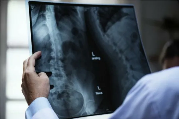
Your typical approach… With some pediatric variations… Pseudosubluxation Ossification Centres OvalContour Loss of Lordosis Soft tissue measurements Pre-dental Space Pseudospread C1
lignment A- Anterior vertebral line B- Posterior vertebral line C- Spinolaminar line D- Spinous processes
Loss of Lordosis Pseudosubluxation
Loss of Lordosis • Distance between spines not > 1.5X adjacent • C1-2 normal up to 10-12mm
Alignment - Odontoid Normal up to 7mm of lateral displacement
one • Oval contour and anterior wedging • Ossification centres
Ossification CentresC1 7 7 3
Ossification CentresC2 6 3 6
Odontoid age 4 Odontoid age 8
So many ossification centres…so little memory… • The spinous process should be fused by 2-3 years • This “wishbone” should fuse with the body by age 6 – can be later in C1 • Extra caution with C2’s late fusing centres: • Base of the dens • Top of the dens
ens 5 mm
Summary • Loss of lordosis, Pseudosubuxation, C1 spread • Oval contour, Ant. Wedging, ossification centres • As in adults • Pre-dental space, Tilt • Changes with age
Case1 Something just doesn’t look right here…
Powers Ratio A D A-B/C-D < 1 C B
A lucency at the physis is not always just the physis Beware the odontoid and all it’s ossification centres
Case Four The absence of a visible neural arch fracture does NOT rule out hangman’s fracture
Summary • Loss of lordosis, Pseudosubuxation, C1 spread • Oval contour, Ant. Wedging, ossification centres • As in adults • Pre-dental space, Tilt • Changes with age