Download
1 / 19
190 likes | 283 Vues
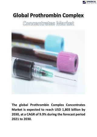
Explore the intricate steps of clotting mechanism, from prothrombin activation to clot formation, and understand the vital role of intrinsic and extrinsic pathways in blood coagulation. Learn about clot retraction, serum, fibrinolysis, anticoagulants like heparin and dicoumarol, and their therapeutic uses.

E N D
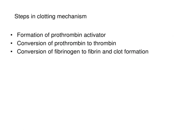
Steps in clotting mechanism • Formation of prothrombin activator • Conversion of prothrombin to thrombin • Conversion of fibrinogen to fibrin and clot formation
Details of coagulation Formation of prothrombin activator, can be by either intrinsic or extrinsic systems. When once the prothrombin activator is formed, the successive steps are common. When clotting occurs in the body, it involves both the intrinsic and extrinsic systems and these systems will complement each other. During the process of clotting, the inactive factors are converted into active forms, and their enzymatic actions produce successive reactions in a cascading manner.
Intrinsic pathway In this, the formation of prothrombin activator, is initiated by platelets. Endothelial damage + Collagen exposure XIIa XII Platelets Kinogen, prekallikrein XIa XI Ca Phospholipids IX IXa VIII, Ca X Xa Phospholipids, Ca Prothrombin activator
Extrinsic pathway Here, formation of prothrombin activator is initiated by tissue thromboplastin. Tissue trauma + Tissue thromboplastin VIIa VII X Xa Phospholipids, Ca,III Prothrombin activator
Special attributes of process of coagulation 1. There is enzyme substrate reaction 2. The system acts as a bioamplifier system. 3. There is cascade reaction. 4. There are +ve and –ve feedback mechanisms involved in the reactions. 5. Ca++ are essential in almost all steps.
clot retraction Freshly formed blood clot is big and soft. As time progresses ( about 30 to 45 mins) , not only the size of clot decreases, the clot becomes firm, a straw colored fluid oozes out. Decrease in size and hardening of clot is due to clot retraction which is brought about by actin-myosin like filaments which are of contractile nature. Decrease in the size of clot can be to the extent of 40 to 60% of the original size.
Purpose of clot retraction Because of the clot retraction the cut ends of the blood vessel come together which adds to hemostasis. When clotting occurs within the blood vessels it is known as thrombosis. This obstructs the blood flow through the blood vessel. Clot retraction facilitates recanalization of the thrombosed veins. When the clot undergoes retraction it becomes firm and prevents thromboembolism. In any condition where the platelet count is reduced, the clot retraction suffers.
What is serum? Serum is the fluid part that is squeezed out during the process of clot retraction. It is identical to plasma in almost all aspects except for the absence of factor number I, II, V, VIII and XIII. Hence it can’t clot. It is used in the laboratory for many of the tests e.g. estimation of serum bilirubin level.
Fibrinolysis Fibrinolysis is a process by which the clot is broken down. It is essential to prevent clogging or blocking of capillary. After the formation of clot, the fibrinolytic system becomes active. which involves many factors that are present in circulation in inactive form and get activated during fibrinolysis.
Anticoagulants Anticoagulants are substances that prevent clotting of blood. They also have important role to play in maintaining the fluidity of blood in circulation. They can be classified into in vivo and in vitro anticoagulants. 1. In vivo anticoagulants are used to prevent coagulation of blood inside the body. Examples of in vivo anticoagulants are heparin and dicoumarol. 2. In vitro anticoagulants are used to prevent coagulation of blood outside the body. Examples of in vitro anticoagulants are heparin, inorganic salts of sodium, potassium and ammonium.
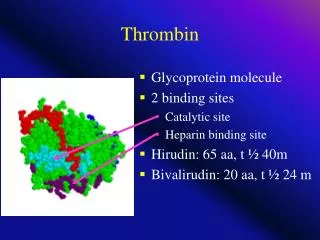
Heparin Heparin is produced by the mast cells & basophils. Mast cells are present in the lining of the blood vessels and lungs. Mechanism of action of heparin are: • Antithrombin action that is, it neutralizes the action of thrombin and prevents conversion of fribrinogen to fibrin. 2. Antithromboplastin action that is, it antagonizes the action of thromboplastin and prevents conversion of prothrombin to thrombin. 3. Prevents aggregation of platelets thereby prevents platelets plug formation. Therapeutically heparin is used for preventing intra vascular thrombosis, during dialysis, myocardial infarction etc.
Dicoumarol Dicoumarol is not present in the body. Exogenous administration is essential. It can be taken orally. It acts as vitamin K antagonist. Vitamin K is essential for the synthesis of coagulation factors II, VII, IX and X by the liver. Therapeutically dicoumarol is used to prevent intra vascular clotting in long standing bedridden patients.
In vitro anticoagulants They are heparin, sodium citrate solution (3.8 %), double oxalate (ammonium and potassium), and EDTA (ethylene diamino tetra acetic acid). Ionic calcium is necessary in many of the steps of coagulation. The inorganic salts will remove the ionic calcium either by precipitating calcium ions as salts (sodium citrate, double oxalate) or as chelating agent of calcium (EDTA). In vitro anticoagulants are used to prevent coagulation of blood outside the body mostly in laboratories for investigation purposes and for storing blood in blood banks.
Test for clotting Bleeding time— It’s the time interval from the oozing of blood till the arrest of bleeding Normal duration 3 to 6 min Clotting time— It’s the time interval from the oozing of blood till the formation of clot Normal duration 3 to 8 min
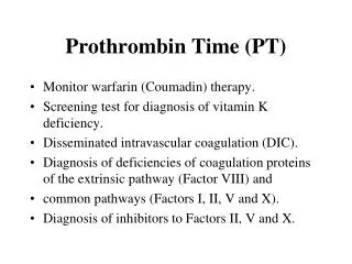
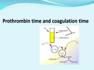
Prothrombin time Prothrombin time is the time interval required to convert prothrombin to thrombin and final clot formation. This measures the extrinsic system involved in blood coagulation. Normal time is about 12 – 16 sec. Prothrombin time increases in liver diseases and forms one of the tests for liver function tests. Prothrombin time is also increased in vitamin K deficiency, as this vitamin is essential for the synthesis of prothrombin
Hemophilia Hemophilia is a disorder of coagulation of blood. It is sex linked inherited disease. Manifests only in males and hence they suffer and females are the carriers. The gene responsible is a recessive gene and is present on the X chromosome. It occurs due to lack of formation of prothrombin activator, so coagulation time is prolonged but BT is normal Deficiency in the formation of prothrombin activator is due to deficiency of factor VIII or IX Hemophilia A: Factor VIII is deficient, about 85% of people with hemophilia are affected by this type. Hemophilia B: Factor IX is deficient here, about 15% of people with hemophilia are affected by this type. Its also called “Christmas disease”
Purpura Here the bleeding time is prolonged but clotting time is normal. Thrombocytopenic purpura– due to deficiency of platelets Thromboesthenic purpura– due to abnormal platelets Idiopathic thrombocytopenic purpura.