Download
1 / 29
370 likes | 748 Vues
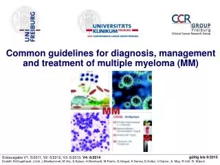
Common guidelines for diagnosis, management and treatment of multiple myeloma (MM). gültig bis 9/2015. Erstausgabe V1: 5/2011, V2: 5/2012, V3: 6/2013, V4: 8/2014

E N D
Common guidelines for diagnosis, management and treatment of multiple myeloma (MM) gültig bis 9/2015 Erstausgabe V1: 5/2011, V2: 5/2012, V3: 6/2013, V4: 8/2014 Erstellt: M.Engelhardt, J.Udi, J.Waldschmdt, M.Vits, S.Kaiser, H.Reinhardt, M.Pantic, G.Herget, K.Henne, E.Kotter, U.Salzer, A. May, R.Voll, R. Wäsch
Allgemeine Hinweise • Eine Therapie im Rahmen von klinischen Studien ist vorrangig zu erwägen, bitte dazu die Details/Auflistung der aktuellen Studien beim Multiplen Myelom im Intranet beachten (Verlinkung) • Jeder neue MM Patient sollte in der Multiplen Myelom Konferenz1 (siehe TOS/Carat+-Tumorboard) vorgestellt werden • Myelom-Datenbank: jeder Patient sollte eine Einverständniserklärung (KMP+Tumordatenbank) erhalten und unterschreiben Für die Inhalte, die richtige Wiedergabe der Beträge und die Richtigkeit der sonstigen Angaben sowie die Aktualität übernimmt das Universitätsklinik Freiburg, Abteilung für Hämatologie, Onkologie und Stammzelltransplantation keine Gewähr. Die Nutzung erfolgt in eigener Verantwortung. 1Montags, 16-17h, Kleiner Hörsaal, UKF 3
MM-Pathway im Blauen Buch Engelhardt, Berger, Duyster, Mertelsmann. The Blue Book. 5. edition. Springer, 2014 4
Diagnostic criteria: MGUS, SMM, symptomatic MM UK Nordic Myeloma Guidelines 2005 + NCCN 2.2014 Engelhardt M. et al. Leuk Lymphoma. 2010 Nov;51(11):2006-11 Engelhardt M. et al. Leuk Lymphoma. 2010 Aug;51(8):1424-43 Dimopoulos M. et al. Blood 117: 4701-5, 2011 Engelhardt M et al. Haematologica 99:232-42, 2014 Dispenzieri A et al. Blood :122:4172-81, 2013 *With conventional radiography accordings to Durie & Salmon With whole-body CT according to Durie & Salmon Plus 10
Risk stratification model for MGUS Predictors of progression of MGUS to myeloma or related disorders: • Size of serum M-protein: Initial Serum M-protein >15g/l • Abnormal Kappa/Lambda free-light chain (FLC) ratio • Type of M-protein: IgG vs. IgA or IgM MGUS Robert A. Kyle and S. Vincent Rajkumar bjh; 134: 573–589, 2006 Bird J. et al. bjh; 147: 22-42, 2009 11
Asymptomatische MM=SMM Algorithm for reclassifying SMM and active MM. *If pts with deletion 17p, t(4;14), and 1q21 gains are included as active MM; this population could account for as many as 30% of SMM pts Merlini G and Palladini G. Eductational ASH 2012 Rajkumar SV et al. Leukemia. doi: 10.1038/leu, 2013 *Dispenzieri A et al. Blood :122: 4172-81, 2013 13
Prognostic factors Tumorburden • Cytokine production Proliferation Genetics • Stage (by Durie&Salmon), IgA-MM, MRI /PET scan • LDH • ß2-MG • Albumin International Staging System • CRP • Plasma cell-labeling-Index (PLI) • Histology: plasmablastic disease Chromosomal aberrations Unfavorable del 17p* t(14;16)* t(14;20)* t(4;14)* 1q gain* >1 chr.aberrations of* non-hyperdiploid Favorable t(11;14 t(6;14) Monosomy 13 Hyperdiploid/Trisomy all others • DNA copy number alteration by CGH/SNP assay • Age, performance status and comorbidities a in presence of concurrent trisomies, should be considerd standard-risk. Bersagel PL et al. Blood 121:884-92, 2013 Rajkumar S.V. et al. Am J Hematol. 88(3):226-35, 2013 Kleber, ...Engelhardt, CLML, 2013 15
Time (years) Risk stratification of cytogenetics - literature review Bergsagel L et al, 2013, Blood, 121:884-892 Boyd KD et al , Leukemia, 2012 Gain 1q21, del(17p13), t(4;14), t(14;16), t(14;20) Rajkumar V, Am. J. Hematol, 2012 Mikhael JR, Mayo Clinic 2013, mSMART Avet-Loiseau H et al, JCO, 2012 1 Moreau P. JCO 2014 2 Bianchi G, Richardson P, Anderson K. JCO 2014 1 Age >55, ß2-MG >5.5 mg/l; t(4;14), del(17p), 1q gain 2 LDH, ISS 3, t(4;14) and/or del17p
Initial investigation/diagnostics in MM FBC: full blood count FISH: fluorescence in situ hybridization * The highest number of plasma cells obtained by either procedure is recorded grey color: optional, e.g. with clinical symptoms. Dimopoulos M. et al. Blood 117: 4701-5, 2011 von de Donk. Haematologica 99:984-96, 2014 Engelhardt M. Haematologica 99:232-42, 2014 NCCN guidelines 2.2104 5
Serum Immunfixation (IF) - MM-Diagnostik: Wissenswertes und sinnvolle Bestimmung Hauptindikationen und Wissenswertes: • Weitere Abklärung scharfe Bande oder „Peak“ in Serum Elektrophorese (SPEP). • Bei normaler SPEP und vorhandenem V.a. MM, Morbus Waldenström (WM), primäre Amyloidose (AL), solitäres oder extramedulläres Plasmozytom. • Essentiell, um monoklonale von polyklonaler Immunglobulin-Erhöhung zu unterscheiden. • Deutlich sensitiver als SPEP (10-50x), Sensitivität SPEP: 1-2 g/L, IFE: 0,1-0,2 g/L) • Zur Bestimmung Schwerketten- und Leichtketten-Typ eines monoklonalen Proteins. • Bestimmt nicht - anders als die SPEP - die Serum Konzentration des M-Proteins (->ZL). Weitere Indikationen und seltene Konstellationen: • Bei Patienten mit MM oder Makroglobulinämie, wenn nach Therapie Bande in SPEP nicht mehr detektierbar ist, z.B. Festlegung CR • Bestimmung und Unterscheidung von biklonalen (2 M-Proteine) und triklonalen (3 M-Proteine) Gammopathien, die in SPEP als einzige Bande oder „Peak“ erscheinen können. • Kann monoklonale k/l Bande nicht IgG, IgA oder IgM oder freien k/l LK zugeordnet werden, muss IF mit anti-IgE + -IgD Antiserum durchgeführt werden. • Frequenz/ Sinnvolle Wiederholung: • Beim Nachweis eines monoklonalen Proteins und Bestimmung des M-Protein Typs mittels IF ist routinemäßige Wiederholung (z.B. alle 1-3 Mon.) der IF nicht notwendig. • Insbes. notwendig zur Dokumentation komplette Remission (CR). • Günstig: genaue Fragestellung, klin. Angaben + ggf. Rü mit Immunologie/Rheumatologie (Drs. Salzer, Udi) Monoklonale Gammopathie vom Typ IgGk (exemplarische SPEP und IF) UpToDate 2014, www.uptodate.com 6
Algorithm for imaging+bone disease management Suspected plasmocytoma or MM Suspected spinal cord compression Soft-tissue mass Whole body CT Urgent MRI scan and appropriate medical management MRI1 and consider biopsy Lytic lesions present Yes Focal lesions >1 Diffuse pattern 0-1 Focal lesions No diffuse pattern Systemic therapy* Observation At risk for fracture? Yes Urgent orthopedic review: consider RTX or operative intervention + * 1 MRI (Orthopädie, UKF) 2 Orthopädie UKF Terpos et al. JCO; 29: 1907-19-15; 2011 Terpos, Zenith Meeting 2012 1,2 Personal communication: PD Dr. Herget / PD Dr. Hauschild, Orthopädie, UKF, Prof. Dr. E. Kotter, Radiologie 9
MM und MGUS: Bildgebende Diagnostik-UKF Allgemein: • Schnittbildverfahren deutlich sensitiver, somit Pariser Schema überlegen • Für Beurteilung Osteodestruktion/Stabilität: Ganzkörper (GK)-CT erforderlich • MRT: für Nachweis diffuser KM-Befall + Weichteilinfiltration (extramedulläres MM) ED, V.a MGUS, smoldering multiple myeloma (SMM) + symptomatisches MM • Bei symptomatischem MM mit V.a. Knochenläsionen: in der Regel GK-CT, bzw. *Nativ-Röntgen symptomatischer Regionen der langen Röhrenknochen/WS-Becken • Bei V.a. extramedulläres MM: zielgerichtetes MRT Low-risk MGUS (Mayo-Risikofaktoren: 0-1) • Radiologische Diagnostik nicht routinemäßig, symptomorientiert • Im Verlauf: GK-CT bei klinischer Indikation, ggf. *Röntgen nativ lange Röhrenknochen/WS SMM, solitäres Plasmozytom und High-risk MGUS (RF nach Mayo 2+3) • GK-CT bei klinischer Indikation, ggf. ergänzend *Röntgen nativ Engelhardt M, Kleber M,... Durie BG. Anticancer Res. 29:4745-50; 2009 Terpos et al. JCO; 29: 1907-19-15; 2011 Hillengass , Delorme, Radiologe.;52:360-5; 2012 van de Donk. Haematologica 99:984-96; 2014 Engelhardt M et al. Haematologica 99: 232-42, 2014 NCCN guidelines 2.2014 Personal communication Prof. Dr. Kotter, Radiologie UKF; PD Dr. G. Herget, Orthopädie, UKF 8
AL-Amyloidose-Diagnostik Bei welchen Patienten? • Prinzipiell kann bei jedem MGUS AL-Amyloidose bestehen; bevorzugt bei: - Paraprotein vom Typ Lambda - Zytogenetik t(11;14) - Erhöhten freie Leichtketten im Serum Klinische Symptome? • Makroglossie, periorbitale Einblutungen, Synkopen, Durchfälle, Herzinsuffizienz, Ödeme, Polyneuropathie Welche Diagnostik ist geeignet? • EKG, NT-Pro-BNP,TropT, Albuminurie, S-FLC • Echokardiografie, Neurologische Untersuchung, Oberbauch-Sonographie/ Gamma-GT Diagnostik zum Amyloidosenachweis: • Fad pad, KMP, Recto-/Gastroskopie (tiefe+serielle Biopsie, wenn möglich) • Niere + Herz (letztere erfolgt aufgrund Risiko seltener, stattdessen typischer Herzechobefund maßgebend) • Histopatholog. Nachweis AL-Amyloidose (vs. z.B. ATTR od. senile Amyloidose) + Paraproteinnachweis vor anti-MM/AL-Amyloidosetherapie 7
Multiples Myelom - Stadieneinteilung International Staging System Durie & Salmon Stadium I ß2-MG < 3,5mg/ml, Albumin 3,5 g/dl Stadium II ß2-MG < 3,5mg/ml, Albumin < 3,5 g/dl oder ß2-MG 3,5-5,5 mg/dl Stadium III ß2-MG > 5,5mg/ml Stadium I Alle folgenden Kriterien müssen erfüllt sein - Hb 10 g/dl - Serumkalzium normal ( 12 mg/dl = 2,75 mmo/l) - Skelett-Röntgen: maximal eine solitäre Läsion - IgG 5g/dl; IgA 3 g/dl - Bence-Jones-Proteinurie 4 g/24h Stadium II - Befunde weder den in Stadium I noch III entsprechend Stadium III Mindestens eines der folgenden Kriterien muss erfüllt sein - Hb 8,5 g/dl - Serumkalzium erhöht (>12mg/dl = >2,75 mmol/l) - Skelett-Röntgen: 2 Osteolysen - IgG >7 g/dl; IgA > 5 g/dl - Bence-Jones-Proteinurie >12 g/24h Subklassifikation A Serumkreatinin < 2 mg/dl B Serumkreatinin 2 mg/dl ß2-MG: ß2-Mikroglobulin 14
UKF-Pathway: Newly diagnosed MM Diagnosis of MM (symptomatic MM) Assessment: age, comorbidities, ISS, cytogenetic, extramedullary disease, bone disease Candidate for autologous stem cell transplantion Yes No DSMM XIII/XIVtrials Recommended initial treatment (6-9 cycles) or VCD VMP Alkylator + steroids + IMIDs: CTD MPT Induction-3-drug regimen 3 x VCD Stem cell harvest High-dose melphalan Additional options: • Bendamustine / prednisone • VMPT-VT • MPR CR/vgPR No risk factors: Cytogentic, ISS I and no renal impairment Consolidation • 2. transplant • Bortezomib • Lenalidomide Maintenance • Lenalidomide • Thalidomide • Bortezomib Maintenance (SD/MR) • Lenalidomide • Thalidomide • Bortezomib No treatment Engelhardt, Berger, Mertelsmann. The Blue Book. 5. edition. Springer, 2014 Ludwig et al. The Oncologist, 17: 592-606, 2012 Palumbo and Roberto,Blood Rev, 2013 Palumbo et al. Haematol., 2012 Engelhardt M et al. Haematologica 2014 17
UKF-Pathway: Relapsed/refractory MM Consider ASCT 2. ASCT for patients in remission > 2 years after first-line transplant allo-SCT may be option for specific patients Frontline treatment with novel agents Yes No Use novel agents Repeat or change frontline treatment Re-treatment feasible after: • Long remission (>6 months) • No toxicity concerns from first line treatment Switch drug class, especially after: • short remission (<6 months) • toxicity concerns from previous line IMiD-based: • Len/dex • Thal/dex • CTD Bortezomib-based: • Borte +/- Dex • VTD • VCD Frontline consisted of: Bortezomib+ IMiD based: • VMPT • VTD IMiD-based (CTD, MPT, RD or Rd) Borte-based (VMP or VD) Consider: clinical trials: VBDD (IIT) or Poma-trial IMiD-based: • Len/dex • Thal/dex • CTD Bortezomib-based: • Borte +/- Dex • VTD • VCD Ludwig et al. The Oncologist, 17: 592-606, 2012 van de Donk et al. Cancer Treat Rev.; 37: 266-283, 2012 Engelhardt M et al. Haematologica 2014 18
BB-protocols for the treatment of MM 5 23 5 8 3 6 total: 50 (2004: 11) Engelhardt, Berger, Mertelsmann. The Blue Book. 5. edition. Springer, 2014 22
Anti-MM systemic treatment options according to age Engelhardt M. et al. Haematologica 2014 Schnerch,...Engelhardt M. IF Onkologie 2014 Kortüm,KM & Stewart AK. Blood 212:893-7, 2013 Engelhardt M et al. RR Cancer Research, Springer, 204 16
CTx-dose reduction in elderly pts or 1.3mg/m2 d1+8 (+15) Palumbo et al. NEJM 364: 1046-60, 2011 23
DSMM XIV(<65y): bRAD vs VRD Induction R maint. until PD A ⅔ 3x bRAD R Mel 200+ ASCT R maint. until PD > VGPR Mel 200+ ASCT Cy R SD R§ < VGPR alloSCT (Treo/Flu) 1-year R maint. ⅓ B 3x VRD § Randomisation between 2nd Mel200 vs allo SCT for all subjects with PR or SD donor search for MRD or MUD Assumption for induction: bRAD 10% CR, VRD 15% CR; Testing on non-inferiority for bRAD, i.e. < 15% „real“ difference A > VGPR post HD-Mel: second Mel200 (Tandem-Mel) to increase PFS from 25 34 Mo. B < VGPR post HD-Mel : allo-SCT to prolong PFS from 31 to 62 Mo. Analysis of molecular response by immunophenotyping/PCR 19
DSMM XIII study (60-75y) R A2 A1 Dosisreduktion bei eGFR<50 R + low Dex R + low Dex R + low Dex R + low Dex R + low Dex R + low Dex MOB MOB MEL 140 R + low Dex R + low Dex MEL 140 R + low Dex until progression Revlimid maintenance 20
VBDD - IIT Freiburg Vorinostat Bortezomib Doxorubicin Dexamethasone d1 d8 d15 d22 repeat: d29 PI: M. Engelhardt 21
MM-Bisphosphonat-Therapie Wer sollte Bisphosphonate erhalten? • Symptomatisches MM oder solitärem Plasmozytom, nicht MGUS Welches Bisphosphonat? • Zoledronsäure 4mg als KI (15 min) alle 4 Wochen oder Pamidronsäure 60-90mg als Infusion über 3-4 Stunden alle 4 Wochen (mit normaler Nierenfunktion) Bisphosphonattherapie bei Niereninsuffizienz (Creatinin-Cl <30ml/min)? • Ø Pamidronat und Zoledronat • Empfehlung für Clodronat: 50-80CrCL (75% DR), 12-50CrCL (50-75% DR), <12 (50% oder Unterbrechung) Applikationsdauer der Bisphosphonate? • Jahr 1 + 2: alle 4 Wochen, ab 3. Jahr gemäß individuellem Remissionsstatus: bei CR 1x jährlich, sonst 1x alle 2-3 Monate; bei Progress 1x monatlich Prophylaxe von Kieferosteonekrosen? • Vor Beginn und im Verlauf einer BP-Therapie alle 6 Monate: Kontrolluntersuchung beim Zahnarzt. • Vor Beginn eines zahnärztlichen Eingriffs (Zahn-Extraktion, Wurzelbehandlung, Kiefer-OP): Unterbrechung BP-Therapie mind. 1 Monat vorher und 3 Monate nachher; prophylaktische Antibiotika-Therapie (z.B. Clindamycin 4 x 300 mg oder Amoxicillin 3x1g über 10 Tage mit Beginn 2 Tage vor dem Eingriff) Terpos et al. Blood.121:3325-8, 2013 Kortüm, Engelhardt, Rasche, Knop, Einsele. Internist 54:963-77, 2013 26
Supportives + monitoring on induction or salvage therapy Mehta J, Cavo M, Singhal S. How I treat elderly patients with myeloma. Blood 116: 2215-23, 2010 Engelhardt. Haematologica 2014 Schnerch,....Engelhardt. IF Onkologie 2014 Engelhardt M. et al. Onkologe 3:217-28, 2014 *Engelhardt, Wäsch, Landgren, Kleber. CLML 14:98-101, 2014 König,...Engelhardt M. Ann hematol 93:479-84, 2014 König,....Engelhardt CLML 13:671-80; 2013 27
Verlaufsdiagnostik nach MM-Therapie (außerhalb von Studien) • Anamnese: Karnofsky-Index, Infektionen und Begleiterkrankungen • Körperlicher Untersuchungsbefund inklusive Größe und Gewicht: Polyneuropathien?, Infektionen? und Schmerzlokalisation? • Laboruntersuchungen (Blut): Blutbild mit Differentialblutbild, Gesamtprotein, Albumin, Kreatinin, Harnstoff, Natrium, Calcium, Kalium, GOT, GPT, g-GT, Bilirubin, LDH, AP, Harnsäure, CRP, ß2-MG, Serum-Elektrophorese mit Immunfixation, freier Leichtkettentest (SFLC), eGFR (MDRD), quantitative Immunglobulin-Bestimmung, Immunfixation • Knochenmark-Diagnostik: bei initialer KMP, keine direkte Verlaufskontrolle nach Therapie (außerhalb von Studien) bei Bestimmbarkeit Paraprotein + damit Remissionsstand wichtig aber zur Remissionsfestlegung vor auto- (+ allo-) SZT • Bildgebung: im Verlauf bei klinischer Indikation Engelhardt. Haematologica 2014 van de Donk. Haematologica 2014 Schnerch,....Engelhardt. IF Onkologie 2014 Engelhardt M. et al. Onkologe 3:217-28, 2014 28
MGUS-Diagnosestellung/Verlaufskontrollen Merlini G and Palladini G. Eductational ASH 2012 Agarwal A. and Ghobrial I. Clin Cancer Res., 9:985-94, 2013 van de Donk et al. Haematologica 99:984-96; 2014 12
Response criteriaEBMT/IBMTR/ABMTR* * EBMT: European Group for Blood and Marrow transplantation; IBMTR: International Bone Marrow Transplant Registry; ABMTR: Autologous Blood and Marrow Transplant Registry. † For patients with non-secretory myeloma only, reduction of plasma cells in the bone marrow by >50% of initial number (partly response) or 25-49% of initial number (minimal response) is required. ‡ In non-secretory myeloma, bone marrow plasma cells should increase by >25% and at least 10% in absolute terms; MRI examination may be helpful in selected patients Rajkumar SV. et al. Blood 117: 4696-4700, 2011 Lonial S & Anderson KC. Leukemia 28:258-68, 2014 24
Response criteriaInternational myeloma working group (IMWG) Rajkumar SV. et al. Blood 117: 4696-4700, 2011 Lonial S & Anderson KC. Leukemia 28:258-68, 2014 25