Download
1 / 54
830 likes | 2.48k Vues
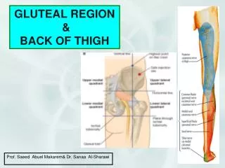
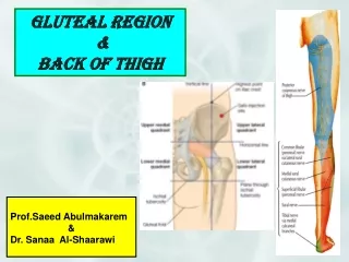
GLUTEAL REGION. Cutaneous nerve supply. Fascia. Ligaments. Muscles. Nerves. Important Arterial Anastomoses. CUTANEOUS NERVES. The gluteal region is divided into four quadrants. Each has its own nerve supply (1) Upper medial

E N D
GLUTEAL REGION • Cutaneous nerve supply. • Fascia. • Ligaments. • Muscles. • Nerves. • Important Arterial Anastomoses.
CUTANEOUSNERVES • The gluteal region is divided into four quadrants. Each has its own nerve supply • (1) Upper medial • Supplied by the posterior rami of the upper three lumbar and the upper three sacral nerves.
CUTANEOUSNERVES • (2) Lower medial: • Posteriorcutaneous nerve of the thigh (S1,2 &3).
CUTANEOUSNERVES • (3) Upper lateral : • Lateral branches of Iliohypogastric (L1) and Subcostal (T12). • (4) Lower lateral : • Lateral cutaneous nerve of the thigh (L2&3).
FASCIA • Superficial Fascia : • Is thick especially in females and contains large quantities of fat to give the characteristic shape of the buttocks. • Deep Fascia : • Continuous inferiorly with the deep fascia of the thigh (fascia lata).
FASCIA • Superiorly it splits to enclose the gluteus maximus muscle . • It covers the gluteus medius and is attached to the iliac crest.
LIGAMENTS • 1. Iliotibial tract. • 2. Sacrotuberous. • 3. Sacrospinous.
ILIOYIBIAL TRACT • It is a vertical thickened band of the deep fascia on the lateral side of the thigh. • Attachment : • Above : tubercle of the iliac crest. • Below: • 1.Lateral condyle of the tibia. • 2.Capsule of the knee joint. 3.Patella.
MUSCLES INSERTED • It is reinforced by fibers from: • 1. Superficial (3/4) of gluteus maximus. • 2. Tensor fascia latae.
FUNCTION • Contraction of these two muscles tighten the tract and it acts as a splint for the knee. • It helps to make the lower limb works as a rigid column.
LIGAMENTS • 2.Sacrotuberous: • It connects the back of the sacrum to the ischial tuberosity. • 2. Sacrospinous : • It connects the back of the sacrum to the ischial spine.
LIGAMENTS • Function : • They stabilize the sacrum and prevent its mobility at the sacroiliac joint by the weight of the vertebral column.
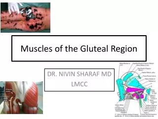
MUSCLES • (1) Three Glutei : • Maximus. • Medius. • Minimus. • (2) Tensor Fascia latae. • (3) Six Lateral Rotators.
THREE GLUTEAL MUSCLES • The gluteus maximus covers the gluteus medius and the (6) lateral rotators. • The gluteus medius covers the gluteus minimus. • The gluteus minimus rests immediately upon the iliac bone.
GLUTEUS MAXIMUS • It is one of the largest, thickest and most powerful muscles of the body. • It has an extensive Origin: • (1) upper part of the ileum behind the posterior gluteal line. • (2) back of the sacrum. • (3) sacrotuberous ligament.
GLUTEUS MAXIMUS • Insertion : • Superficial¾ to the iliotibial tract. • Deep¼ to the gluteal tuberosity of the femur. • Nerve supply : • Inferior gluteal nerve.
ACTION • (1) It is the main extensor of the hip. • It is used only when the thigh has to be extended with Force : • (a) Rising from a sitting position. • (b) Climbing a hill. • C. Running. • It is not used in walking on a level.
ACTION • (2) A powerful lateral rotator of the hip (when the thigh is extended) • (3) Its contraction makes the iliotibial tract tense .
INTRAMUSCULAR INJECTION • The great thickness of gluteus maximus makes it ideal for intramuscular injection. • The injection should be given on the upper outer quadrant of the buttock to avoid injury of the following nerves: sciatic, tibial, common peronealand inferior gluteal.
BURSAE RELATED • Three bursae lie under the gluteus maximus : • 1. A bursa between the lower edge of the muscle and the ischial tuberosity. • 2. A large bursa separating the muscle from the greater trochanter. • 3. A large bursa between the aponeurotic part of the muscle and vastuslateralis.
GLUTEUS MEDIUS • It is a large thick powerful fan shaped muscle. • Origin : • A large area between the iliac crest and the middle gluteal line.
GLUTEUS MEDIUS • Insertion : • In the postero superior angle of the greater trochanter and the oblique line on its lateral surface.
GLUTEUS MEDIUS • Nerve supply : • Superior gluteal nerve. • Action : • 1.Abduction of the thigh. • 2.Medial rotation of the thigh (anterior fibers ).
GLUTEUS MEDIUS • In standing, if the support of one limb is suddenly removed, the gluteus medius of the other side contracts to prevent the pelvis from falling on the unsupported side. • The alternative action of the gluteus medius on both sides is responsible for keeping the pelvis level during walking. • Without the gluteus medius of both sides the gait becomes rolling on a broad base.
GLUTEUS MINIMUS • It is a fan shaped muscle that lies deep to the anterior part of gluteus medius. • Origin : • From the ilium between the middle and inferior gluteal lines.
GLUTEUS MINIMUS • Insertion : • Anterior surface of greater trochanter. • Nerve supply : • Superior gluteal nerve. • Action : • same as gluteus medius.
TENSOR FASCIA LATAE • It is a short thick muscle that lies at the junction of the gluteal region and the upper part of the front of the thigh. • Origin : • From the extreme anterior part of the iliac crest (just behind the anterior superior iliac spine).
TENSOR FASCIA LATAE • Insertion : • In the iliotibial tract. • Nerve supply: • Superior gluteal nerve.
TENSOR FASCIA LATAE • Action : • It tightens the knee so that in walking the knee can take the weight of the body while the other foot is off the ground. • The extension of the knee is made through tightening of the iliotibial tact with the help of gluteus maximus.
SIX LATERAL ROTATORS • Piriformis. • Obturator internus. • Gemelli (superior and inferior ). • Obturator externus. • Quadratus femoris.
SIX LATERAL ROTATORS • These muscles pass behind the hip joint. • They lie below and behind gluteus minimus. • They are covered by gluteus maximus.
ORIGIN • 1. Piriformis • Inside the pelvis mainly from the middle (3) pieces of the anterior surface of the sacrum. • It leaves the pelvis through the GSF which it fills almost completely.
ORIGIN • 2. Obturato Internus: • Inside the pelvis from the obturator membrane and the surrounding bones. • It leaves the pelvis through the LSF.
ORIGIN • 3 & 4. Gemellus superior andGemellus inferior : • From the upper and lower margins of the LS Notch respectively. • They are inserted into the tendon of obturator internus. • 5. Quadratus femoris: • From the ischial tuberosity.
SIX LATERAL ROTATORS • Insertion : • The tendons are inserted into the greater trochanter. • Action : • Lateral rotation of the hip joint.
SIX LATERAL ROTATORS • Nerve supply : • Each of these muscles (Except obturator externus) has its own nerve supply from the sacral plexus or from the sacral nerves.
ORIGIN • 6. Obturator externus • Outside the pelvis from the obturator membrane and the medial and lower margins of the obturator foramen. • It is inserted in the trochanteric fossa.
NERVES • 1. Sciatic nerve. • 2. Posterior cutaneous nerve of thigh. • 3. Superior and Inferior Gluteal nerves. • 4. Nerve to Quadratus Femoris. • 5. Nerve to Obturator Internus. • 6. Pudendal nerve.
SCIATIC NERVE • It is the largest nerve in the body. • It is the larger of the two terminal branches of the sacral plexus. • It is flat and broad near its origin and becomes rounded downwards.
SCIATIC NERVE • It is composed of two components : • (a) Tibial nerve : • It arises from the ventral divisions of all components of the sacral plexus. • (b) Common peroneal verve : • It arises from the dorsal divisions of all components of the sacral plexus.
COURSE • It leaves the pelvis through the lower part of the GSF below the piriformis. • As it descends it lies on: • 1.Root of ischial spine. • 2.Gemelli. • 3.Obturator internus. • 4.Quadratus femoris. • 5.Adductor magnus.
COURSE • It is related posteriorlyto : • 1.Posterior cutaneous nerve of the thigh. • 2.Gluteus maximus. • It enters the back of the thigh by passing deep to the long head of biceps femoris. • Branches: • Usually it has No branches in the gluteal region.
TERMINATION • About the middle of the thigh it divides into its terminal branches :Tibial and Common Peroneal.
POSTERIOR CUTANEOUS N. OF THE THIGH • ORIGIN : • Sacral plexus. • COURSE : • It leaves the pelvis through the lower part of the GSF below piriformis. • It descends on the posterior surface of the sciatic nerve till the popliteal fossa.
POSTERIOR CUTANEOUS N. OF THE THIGH • BRANCHES : • 1.Gluteal : to the lower medial quadrant of the buttock. • 2.Perineal : to the skin of the back of scrotum (or labia majora). • 3.Cutaneous : to the back of the thigh and upper leg.
SUPERIOR GLUTEAL NERVE • ORIGIN : • From the sacral plexus (L4,5 & S1). • COURSE : • It leaves the pelvis through the upper part of the greater sciatic foramen above piriformis. • It passes between gluteus medius and minimus.
SUPERIOR GLUTEAL NERVE • BRANCHES • It gives motor supply to: • 1. Gluteus medius. • 2. Gluteus minimus. • 3. Tensor fascia latae.
INFERIORGLUTEAL NERVE • Origin: • Sacral plexus (L5, S1 &S2). • Course : • It leaves the pelvis through the lower part of the (GSF) below piriformis. • Branches: • It gives motor supply to : • Gluteus maximus.
NERVE TOQUADRATUS FEMORISN • It arises from the sacral plexus. • It leaves the pelvis through the lower part of the GSF below piriformis. • It supplies : • (1) Quadratus femoris. • (2)Inferior gemillus.
PUDENDAL N. & N. TO OBTURATOR INTERNUS • Both arise from the sacral plexus. • They Leave the pelvis through the lower part of GSF below piriformis. • They reenter the pelvis through the LSF with the pudendal vessels by crossing the ischial spine.