Download
1 / 15
230 likes | 1.46k Vues
ANTERIOR & MEDIAL COMPARTMENTS OF THIGH. Dr. Ahmed Fathalla Ibrahim & Dr. Zeenat Zaidi. OBJECTIVES. At the end of the lecture, students should be able to: List the name of muscles of anterior compartment of thigh.

E N D
ANTERIOR & MEDIAL COMPARTMENTS OF THIGH Dr. Ahmed Fathalla Ibrahim & Dr. ZeenatZaidi
OBJECTIVES At the end of the lecture, students should be able to: • List the name of muscles of anterior compartment of thigh. • Describe the anatomy of muscles of anterior compartment of thigh regarding: origin, insertion, nerve supply and actions. • List the name of muscles of medial compartment of thigh. • Describe the anatomy of muscles of medial compartment of thigh regarding: origin, insertion, nerve supply and actions. • Describe the location, boundaries and contents of femoral triangle & adductor canal
The thigh is divided into 3 compartments by 3 intermuscular septa (extending from deep fascia into femur) • Anterior Compartment • Extensors of knee: • Quadriceps femoris • Flexors of hip: • 1. Sartorius • 2. Pectineus • 3. psoas major • 4. Iliacus • Nerve supply: • Femoral nerve • Medial Compartment • Adductors of hip: • 1. Adductor longus • 2. Adductor brevis • 3. Adductor magnus • (adductor part) • 4. Gracilis • Nerve supply: • Obturator nerve • Posterior Compartment • Flexors of knee & extensors of hip: • Hamstrings • Nerve supply: • Sciatic nerve
Anterior Compartment of Thigh • Contains the: • Flexor of the hip: • Sartorius • Pectineus • psoas major • Iliacus • Extensors of knee (Quadriceps femoris): • Rectus femoris • Vastuslateralis • Vastusmedialis • Vastusintermedius (deep to rectus femoris) • Nerve supply: Femoral nerve 4 3 2 1 2 1 4 VastusIntermedius (deep to rectus femoris) 3
Sartorius ORIGIN Anterior superior iliac spine INSERTION Upper part of medial surface of tibia ACTION (TAILOR’S POSITION) • Flexion, abduction & lateral rotation of hip joint • Flexion of knee joint S
Pectineus ORIGIN: Superior pubic ramus INSERTION: Back of femur (below lesser trochanter) ACTION: • Flexion & adduction of hip joint
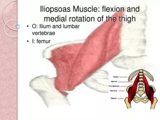
Iliacus & Psoas major (Iliopsoas) ORIGIN: Psoas major: T12 & lumbar vertebrae • Iliacus: Iliac fossa INSERTION: Lesser trochanter of femur ACTION: Flexion of hip joint In less than 50 percent of humans the psoas major is accompanied by the psoas minor. It is located in front of the psoas major muscle.
Quadriceps Femoris ORIGIN: Rectus femoris: Anterior inferior iliac spine Vastusintermedius: Front of shaft of femur Vastusmedialis: Posterior border of femur Vastuslateralis: Posterior border of femur
Quadriceps Femoris INSERTION: Into PATELLA (Patella is a sesamoid bone) From patella into TUBEROSITY OF TIBIA through Ligamentum Patellae (Patellar Ligament) ACTION: • Extension of knee joint
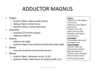
Medial Compartment of Thigh MUSCLES: Adductor longus Adductor brevis Adductor magnus (Adductor portion) Gracilis ACTION: ADDUCTION OF HIP JOINT N.B.: Gracilis also flexes knee joint NERVE SUPPLY: OBTURATOR NERVE 1 2 2 1 4 3 3 4 Adductor magnus (Adductor portion) Adductor magnus (Hamstring portions)
Origin • Inferior pubic ramus • Ischialramus • Body of pubis • Body of pubis • Inferior pubic ramus Adductor part Hamstring hiatus Adductor hiatus Adductor magnus (adductor portion) Gracilis Adductor longus Adductor brevis Insertion • Upper part of medial • surface of tibia • (behind sartorius) • Posterior border of femur (Linea Aspera)
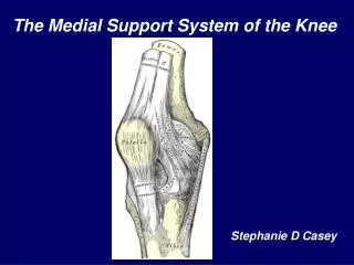
Femoral Triangle: Location & Boundaries • It is a deep hollow in the Upper third of front of thigh inferior to the inguinal ligament Boundaries • Base: Inguinal ligament • Medial: Medial border of the adductor longus muscle • Lateral: Medial border of the sartorius muscle • Floor: (from media to lateral) • adductor longus • Pectineus • Psoas major • Iliacus • Roof: Skin, superficial & deep fascia. Iliopsoas Pectineus
Femoral Triangle: Contents From lateral to medial: • Femoral nerve & its branches • Femora artery • Femoral vein • Lymphatic vessels and some deep inguinal lymph nodes • The femoral artery, femoral vein and the lymph nodes lies within the fascial envelope, the Femoral sheath,occupying its lateral, intermediate and medial compartments respectively. The medial compartment is called the femoral canal. Femoral sheath
DEFINITION: an aponeurotic tunnel for femoral artery & vein SITE: In middle third of front of thigh deep to sartorius EXTENT: From apex of femoral triangle to adductor hiatus BOUNDARIES: Roof (Anterior): Sartorius (medially) and vastusmedialis (laterally) Floor (Posterior): Adductor longus & magnus ADDUCTOR CANAL(Subsartorial/Hunter’s canal) Adductor hiatus